Comparative Assessment of Clinical Effectiveness and Patient Acceptability of Hawley and Vacuum-Formed Retainers- An Analytical Study
Comparative Assessment of Clinical Effectiveness and Patient Acceptability of Hawley and Vacuum-Formed Retainers- An Analytical Study
Dr. Preeti Bhattacharya1, Dr. Abhishek Singh2*, Dr. Anil Kumar Chandna3, Dr. Ankur Gupta4, Dr. Ravi Bhandari5, Dr. Resham Irshad6
Dr. Preeti Bhattacharya - Head of the Department
Dr. Abhishek Singh - Post graduate studentIntern Medical officer at Vydehi Institute of Medical Sciences and Research Centre , Bengaluru, Karnataka, India"
Dr. Anil Kumar Chandna - Director of PG studies
Dr. Ankur Gupta - Professor
Dr. Ravi Bhandari - Professor
Dr. Resham Irshad - Senior Lecturer
Workplace: Institute of Dental Sciences, Bareilly, India
*Correspondence to: Dr. Abhishek Singh, Post Graduate student, Department of Orthodontics and Dentofacial Orthopaedics, Institute of Dental Sciences, Near Suresh Sharma Nagar, Pilibhit bypass road, Bareilly. Pin Code 243006
Copyright.
© 2025 Dr. Abhishek Singh This is an open access article distributed under the Creative Commons Attribution License, which permits unrestricted use, distribution, and reproduction in any medium, provided the original work is properly cited.
Received: 21 Aug 2025
Published: 18 Sep 2025
DOI: https://doi.org/10.5281/zenodo.17181190
ABSTRACT
Background: Post-orthodontic retention is critical to prevent relapse and ensure long-term treatment stability. Among removable retainers, Hawley Retainers (HR) and Vacuum-Formed Retainers (VFR) are the most commonly prescribed.
Aim: To evaluate and compare the clinical effectiveness and patient acceptance with Hawley retainers and Vacuum-formed retainers.
Material and Methods: A total of 45 patients who completed fixed orthodontic treatment were randomly assigned to either HR (Group I) or VFR (Group II). Retainers were worn full-time for 6 months and at night for 3 months. Data were collected at debonding (T0), 3 months (T1), and 9 months (T2). Parameters assessed included archwidth changes (using dental casts), occlusal contacts (using Aluwax impressions), and patient acceptability (using a 10-point Visual Analogue Scale).
Results: Arch Width: Significant intergroup differences were found, with Group II showing greater archwidth changes . Occlusal Contacts: Both groups showed an increase in anterior, posterior, and total occlusal contacts from T0 to T1; however, Group I demonstrated a significantly increase in contact points. Patient Acceptance: Group II shows statistically significant differences in appearance, speech, and self-esteem. Other parameters showed non-significant differences but trend was in favor of Group II.
Conclusion: Both HR and VFR are clinically effective in maintaining orthodontic outcomes. HR promotes better occlusal settling, whereas VFR are more favored by patients for aesthetics and comfort. Clinical choice should consider both functional stability and patient preference.
Keywords: Hawley, Essix, Retainer, Retention Techniques, VFR.
Comparative Assessment of Clinical Effectiveness and Patient Acceptability of Hawley and Vacuum-Formed Retainers- An Analytical Study
Background
The success of orthodontic treatment mainly depends on retaining the teeth in the corrected position after the debonding appointment as they are potentially unstable therefore retention is necessary1. Retention is maintained through orthodontic retainers, which can either be fixed or removable. Removable retainers which are commonly used are Hawley retainers (HR) and vacuum formed retainers (VFR).2,3 HR have been a reliable and effective option for removable orthodontic retention for more than a century. Since the advent of VFR in 1971, it has become increasingly popular. Successful outcome of any retention device depends on its ability to maintain archform, preventing undesirable tooth shifting while allowing required settling.4 Effectiveness also depends on patient’s compliance which in turn again linked to its acceptability.
A study done by Tarman ke, et al. aimed to compare patient acceptance and satisfaction with two types of removable retainers, concluded that VFR were preferred for speech comfort, while no substantial differences were found between the two retainers in other aspects of patient perception.5
Kalaydzhieva M, et al. evaluated the effectiveness of Hawley retainers, Vacuum-formed retainers and fixed retainers in maintaining dental arch dimensions and tooth alignment and found that Vacuum-formed retainers were superior in maintaining maxillary anterior alignment, while Hawley retainers preserved maxillary arch length. Both types of retainers showed no significant difference in maintaining transverse dimensions, intercanine width, interpremolar width and intermolar width.6
The objective of this study was to compare archwidth and how occlusal contacts in maximum intercuspation change over time during the post-retention period, as well as to assess patient compliance in individuals wearing HR and those wearing VFR.
Materials and Methods
Study Design and Population
The study was conducted in the Department of Orthodontics, Institute of Dental Sciences, Bareilly. A total of 45 subjects who completed fixed appliance orthodontic treatment were included. Ethical clearance and informed consent were obtained prior to study commencement.
Patients included in the study had not undergone any prior orthodontic treatment other than that which was provided at the Institute of Dental Science. Additionally, all selected patients were treated using fixed orthodontic appliances in both jaws, following the 0.022-inch slot MBT prescription.
Patients were excluded from the study if they had received single arch or sectional fixed appliance treatment, had undergone Rapid Maxillary Expansion, or required prosthetic treatment for missing teeth. Additionally, individuals with learning disabilities, cleft lip and palate, poor periodontal status, or temporomandibular disorders were not included in the study.
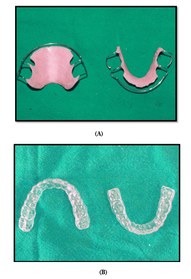
Participants were then randomly allocated to receive either Hawley Retainer (HR) or Vacuum-Formed Retainers (VFR) in both arches [Figure 1]. Based on the type of retainer provided, they were divided into two groups:
Group - I: Subjects who were given Hawley Retainers
Group - II: Subjects who were given Vacuum Formed Retainers
Figure 1 - (A) Hawley retainer; (B) Vacuum- formed retainer
Intervention and Procedure
Hawley Retainers were fabricated using acrylic resin and 21-gauge stainless steel wire. In patients who had undergone premolar extraction, a long labial bow extended from second premolar of 1st quadrant to second premolar of 2nd quadrant and C clasps on the first molars were incorporated. For non-extraction cases, a short canine-to-canine labial bow with Adams clasps on the first molars were used. The labial bow was adjusted to make light contact with the labial surfaces of the incisors.
Vacuum-Formed Retainers (VFR) were made from 1 mm thick polyvinyl siloxane sheets. These were trimmed to extend 1–2 mm over the labial gingiva and 3–4 mm over the palatal or lingual gingiva, covering the occlusal surfaces of all teeth, including the most distally erupted molar.
Retainers were delivered and fitted within 24 hours of debonding. Participants were instructed to wear their retainers full-time for the first 6 months, followed by nighttime wear thereafter. Retainers were advised to be removed only during meals, cleaning, and drinking hot beverages. Fit and adaptation were checked during each follow-up appointment.
Collection of Data
Dental Casts
The maxillary and mandibular dental casts were obtained from all patients at 3 points in time. The first set of records were taken at T0 (at the time of debonding); the second set at T1 (3 months after delivery of retainers) and third set at T2 (9 months after delivery of retainers).
Occlusal Records
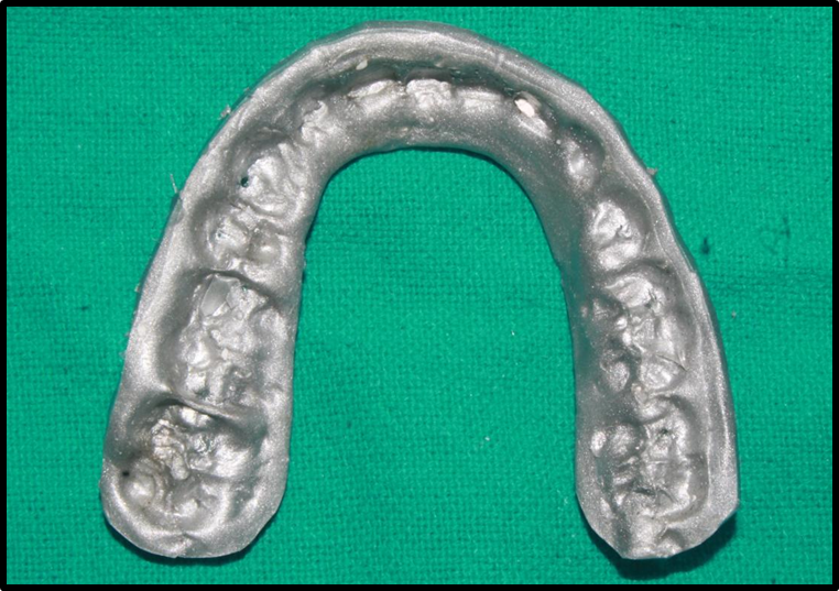
Occlusal records were obtained from all patients at two points of time i.e immediately after debonding (T0) and after three months (T1), using Aluwax bite registration. Subjects were made to sit upright, and softened Aluwax was placed on the mandibular occlusal surfaces. Patients were guided to bite in maximum intercuspation firmly and maintain the position for about two minutes. In cases where the initial registration was inadequate due to insufficient material or improper placement, a second bite registration was performed.[Figure 2]
Figure 2 - Occlusal record using Aluwax
Patient Acceptability Assessment
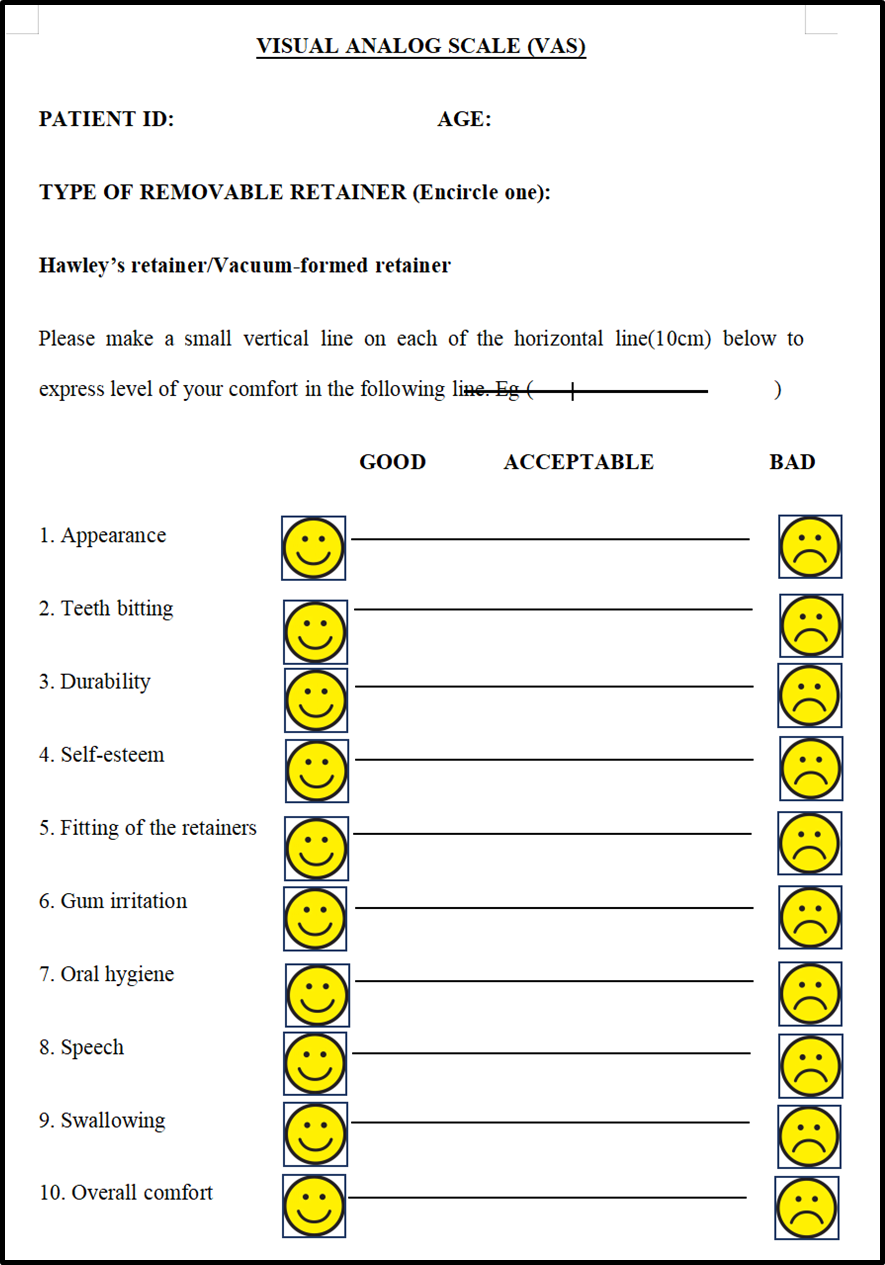
Subjects were evaluated three months after retainer placement (T1) using a 10-cm Visual Analogue Scale (VAS). It consisted of 10 questions related to retainer acceptance. Both oral and written instructions were provided to ensure proper completion of the questionnaire, which ranged from 0 (most favorable response) to 10 (least favorable). The assessments were conducted in the presence of the treating doctor. [Figure 3]
Figure 3 - Visual Analogue Scale (VAS)
Analysis of Data
Measurement on Dental cast
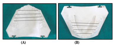
Maxillary and mandibular dental casts were obtained for each subject, and transverse arch dimensions were recorded using an electronic digital caliper (Mitutoyo Digital Vernier Caliper 0–150 mm) with a precision of 0.01 mm . For the maxillary cast, four measurements were taken: Intercanine Width (ICW), defined as the distance between the cusp tips of the right and left canines; Interpremolar Width (IPMW), measured between the buccal cusp tips of the second premolars on both sides; Interfirst Molar Width 1 (IFMW1), the distance between the mesiobuccal cusp tips of the first molars on the right and left quadrants; and Interfirst Molar Width 2 (IFMW2), the distance between the distobuccal cusp tips of the first molars [Figure 4]. Similarly, for the mandibular cast, the same parameters were recorded.
Figure 4: (A) & (B) Shows archwidth measurement in maxillary and mandibular arch respectively
Measurement on occlusal records
Interocclusal records were analyzed on a radiographic viewing screen in a dark room to identify areas of perforation and translucent zones without perforation. Contact points were assessed for the first molars, second premolars, canines, lateral incisors, and central incisors. Anterior contacts included central incisors, lateral incisors, and canines, while posterior contacts included the first molars and second premolars. The total occlusal contacts were determined by summing the anterior and posterior contact counts.
Measureme