Minimal Invasive Approaches to Maxillary Atrophy: Two Case Reports Featuring One-Piece Tissue Level Implants and Fixed Screw Retained Bridges
Minimal Invasive Approaches to Maxillary Atrophy: Two Case Reports Featuring One-Piece Tissue Level Implants and Fixed Screw Retained Bridges
Dr Jimoh Olubanwo Agbaje 1, Dr. Henri Diederich *2, Dr Mohamed Seghir Babouche 3
- OMFS-IMPATH Research Group, Department of Imaging and Pathology, Faculty of Medicine, Catholic University Leuven, Belgium.
- 114 av de la Faiencerie, L- 1511 Luxembourg.
- Cité du 1 novembre 1954, El Eulma, Setif, Algeria.
*Correspondence to: Dr. Henri Diederich, Doctor in Dental Medicine, 114 av de la Faiencerie,
L- 1511 Luxembourg.
Copyright
© 2023 Dr. Henri Diederich. This is an open access article distributed under the Creative Commons Attribution License, which permits unrestricted use, distribution, and reproduction in any medium, provided the original work is properly cited.
Received: 25 October 2023
Published: 30 October 2023
Abstract
Atrophy of the maxilla poses significant challenges to dental rehabilitation, particularly in the context of dental implant placement. There exist numerous approaches to manage such scenarios, ranging from invasive techniques such as zygomatic implants, sinus grafting, and bone grafting, to more minimal invasive methods utilizing pterygoid implants and the transnasal approach. The latter techniques are particularly interesting due to their potential for early loading, where patients can receive a fixed rehabilitation within 2-3 weeks post-operatively. This article aims to present two case reports showcasing the clinical use of one-piece tissue level implants and fixed screw retained bridges in the management of maxillary atrophy.
Keywords: Rehabilitation, full arch implant prosthesis, One-piece Implants, Pterygoid implants, transnasal approach, One Piece Tissue level implants
Minimal Invasive Approaches to Maxillary Atrophy: Two Case Reports Featuring One-Piece Tissue Level Implants and Fixed Screw Retained Bridges
Introduction
The rehabilitation of an atrophied maxilla presents a complex, multi-faceted challenge for the dental professional1- 3. A variety of strategies have been developed over the years, each with its unique set of benefits and drawbacks. The more invasive techniques, while often effective, involve a greater level of surgical trauma, higher cost, longer recovery times, and a higher risk of complications 4- 7. On the other hand, minimal invasive approaches offer several advantages, including shorter healing periods and less surgical morbidity, albeit with their own set of challenges 8 -11. Among these, the use of pterygoid implants and the transnasal approach, when applied with specific implants, may allow for early loading, providing patients with a functional, aesthetic, and stable oral rehabilitation within 2-3 weeks 5,10. This paper present two cases that illustrate the application and effectiveness of these minimally invasive strategies.
Case 1
This case presents a report of early loading on One Piece tissue level implants when Pterygoid and nasal approach were used.
The patient a lady of 53 had advanced periodontitis which made her came several times to the clinic for emergency treatment of pain. At a point patient wanted a permanent solution to this problem.
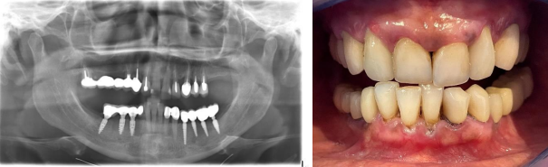
After consultation, plan was made as follows: to extract remaining teeth in the maxilla and mandible, the existing crowns on implants in the mandible were to be changed to have the same color for all the teeth (Vita B1) in the mouth Fig 1.
Figure 1 shows the panoramic radiograph and clinical photo at presentation.
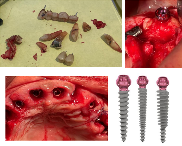
Surgery was done in one session under local anesthesia, teeth in the maxilla and mandible were extracted and implants were placed.
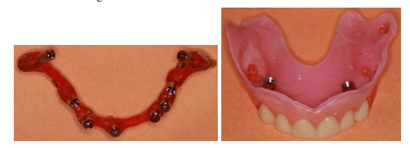
In the maxilla Compressive Implants P 3.5/20 mm (ROOTT) were placed in the extraction sockets and at position 18 and 28 for high primary stability. The high primary stability is achieved due to the specific characteristics of these implants (Fig 2). The long thin part guides the implant during insertion while the larger part with compressive threads compresses the bone which results in bone corticalization.
The implants were inserted to bone level so that the head can be at tissue level. The same procedure was done in the anterior part of mandible.
Figure 2: Surgical procedure and implant placement in the maxilla
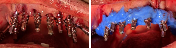
After surgery impressions were taken in the maxilla and mandible, temporary bridges were then made at chairside
Figure 3: Impression procedure
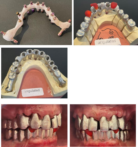
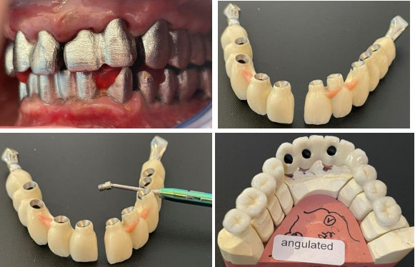
On the second appointment the verification key was tried in, bite relation as well as aesthetical outcome were checked and validated. Ten days later frame was tried in Figure 4.
The metal ceramic bridges were placed in the mandible and maxilla10 days after frame try-in.
The bridge in the maxilla is in 2 parts and screw retained, here angulated screw channels were used in order to get perfect fit. In the mandible the bridge is screw and cement retained.
Figure 4: Laboratory steps: frame try-in and metal-ceramic bridge
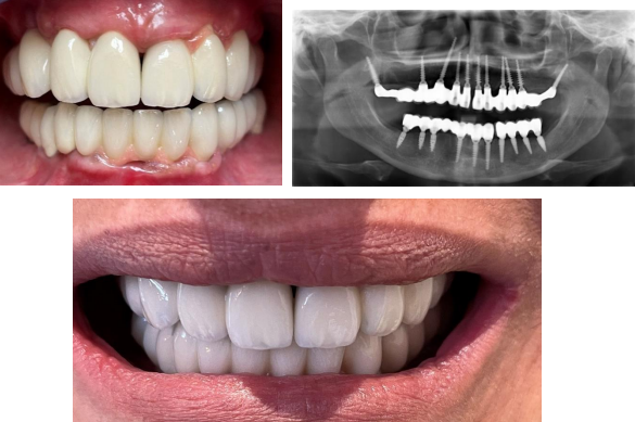
Three weeks after initial consultation, patient´s mouth was rehabilitated with definitive metal ceramic bridges. This was made possible due to the achieved bicortical anchorage in the anterior part and a strong cortical anchorage at the pterygoid plates (75 N/cm) in the maxilla, these anchorage ensured high primary stability which encourages immediate loading. Fig 5
Figure 5: Clinical photograph and the panoramic radiograph of patient and after bridge delivery, day of placement
Case II
The patient, a lady of 56 years old came to clinic with periodontal problems, she desired a solution and oral rehabilitation in a minimal invasive way.
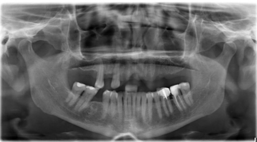
After intra oral examination and radiological reviews (panoramic radiograph and cone beam computer tomography scan) It was observed that the width and height of alveolar bone is not enough for classic implant placement, as a result the choice of appropriate implant is important fig 6.
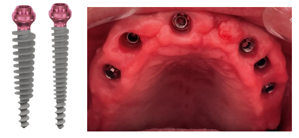
With bone width of 5 to 6 mm One piece Tissue level implants (ROOTT, TRATE AG) were the implants of choice.
Figure 6
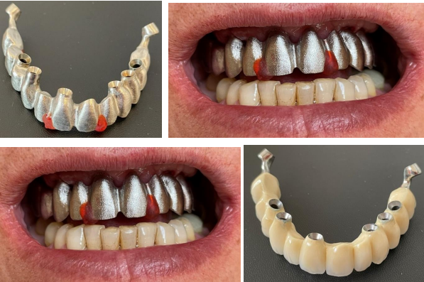
On the first appointment all teeth in the maxilla were extracted, and implants were placed, under local anesthesia. Pterygoid Implants C 3.5/20 mm were placed in position 18 and 28 to get a high primary stability, same implants were used at the extraction’s sockets and in the rest of the maxilla figure 7
After the surgery the impression was taken immediately with screwed transfers and a temporary bridge was made at chairside. Fig 8
Figure 8: Laboratory steps - screwed transfers and aesthetical trial.
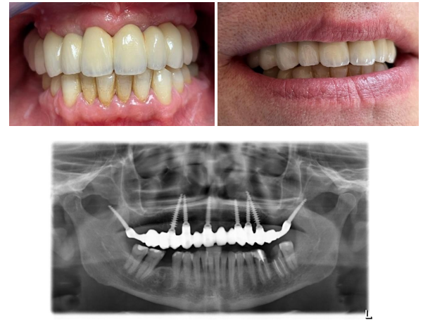
The transfer key was tried in on the second appointment to check the impression, an aesthetical trial was also done. After validation third appointment was fixed to try in the frame Fig 9
A metal ceramic bridge was fabricated after frame try-in. Three weeks after the beginning of the treatment, the gum has healed nicely, bridge was screw-retained with angulated screw channels so as to get a perfect fit fig 10.
Figure 10
(please click here to view complete article with images)
Discussion
This article presents two case reports that showcase the clinical use of One-Piece tissue level implants and fixed screw retained bridges in the management of maxillary atrophy. Here a minimal invasive method of transnasal approach and Pterygoid implant placement show that an early loading with specific implants ensuring a high primary stability.
The transnasal approach with pterygoid implants techniques are particularly interesting due to their potential for early loading, where patients can receive a fixed rehabilitation within 2-3 weeks post-operatively.
References
1. Ali SA, Karthigeyan S, Deivanai M, Kumar A. Implant rehabilitation for atrophic maxilla: a review. J Indian Prosthodont Soc 2014; 14:196-207
2. Mericske-Stern RD, Taylor TD, Belser U. Management of the edentulous patient. Clin Oral Implants Res 2000; 11 Suppl 1:108-25.:108-125
3. Chan MF, Narhi TO, de BC, Kalk W. Treatment of the atrophic edentulous maxilla with implant-supported overdentures: a review of the literature. Int J Prosthodont 1998; 11:7-15
4. Won?Bae ; Kim, Young?Jin ; Kang, Kyung Lhi ; Lim, Hyun?Chang ; Han, Ji?Young Hoboken, Long?term outcomes of the implants accidentally protruding into nasal cavity extended to posterior maxilla due to inferior meatus pneumatization Park, Clinical implant dentistry and related research, 2020, Vol.22 (1), p.105-111
5. Mazor, Ziv ; Lorean, Adi ; Mijiritsky, Eitan ; Levin, Liran Nasal Floor ElevationCombined with Dental Implant Placement Clinical implant dentistry and related research, 2012, Vol.14 (5), p.768-771, Article 768
6. Candel E, Penarrocha D, Penarrocha M. Rehabilitation of the atrophic posterior maxilla with pterygoid implants: a review. J Oral Implantol 2012; 38 Spec No:461-6. doi: 10.1563/AAID-JOI-D-10-00200. Epub@2011 May 13.:461-466
7. Lorean, Adi ; Mazor, Ziv ; Barbu, Horia ; Mijiritsky, Eitan ; Levin, Liran Nasal Floor Elevation Combined with Dental Implant Placement: A Long-Term Report of up to 86 Months The International journal of oral and maxillofacial implants, 2014, Vol.29 (3), p.705-708, Article 705
8. Agbaje JO, Diederich H. Minimal Invasive Concept for the Rehabilitation of Edentulous Jaw with One-piece Implants. International Journal of Case Reports & Short Reviews 2018; 4:028-030
9. Agbaje JO, Diederich H. Cortically Fixed at Once Implants for the Treatment of the Atrophic Maxilla - A Case Report. Adv Dent & Oral Health 2019; 1:001-005
10. Agbaje JO, Meeus J, Vrielinc L, Diederich H. Rehabilitation of Atrophic Maxilla using Pterygoid Implants: Case Reports. Modern Approaches in Dentistry and Oral Health Care 2018; 1:1-6
11. Candel E, Penarrocha D, Penarrocha M. Rehabilitation of the atrophic posterior maxilla with pterygoid implants: a review. J Oral Implantol 2012; 38 Spec No:461-6. doi: 10.1563/AAID-JOI-D-10-00200. Epub@2011 May 13.:461-466.