A Rare Presentation of Cervical Tardive Dystonia in an Antenatal Patient
A Rare Presentation of Cervical Tardive Dystonia in an Antenatal Patient
Dr Charmi Shah, M.S. Obgy *1, Dr Dipti Jain. M.S. Obgy 2, Dr Manoj Kargathiya. M.D. 3
1.Laproscopic Surgeon.
2.Fellowship in Gynae Oncology.
3.Anaesthetist
*Correspondence to: Dr Charmi Shah, M.S. Obgy, Laproscopic Surgeon.
Copyright
© 2023 Dr Charmi Shah. This is an open access article distributed under the Creative Commons Attribution License, which permits unrestricted use, distribution, and reproduction in any medium, provided the original work is properly cited.
Received: 14 November 2023
Published: 30 November 2023
Abstract
An antenatal patient of 24 years experienced sudden jerky and continuous involuntary movements in her cervical spine during her fourth month. Despite these movements, she remained conscious and oriented, with stable vital signs. Her medical history included normal MRI brain with spine, normal ANA by IF, TSH, B2 glycoprotein, serum copper, ceruloplasmin, and normal ASO titre. Upon further examination, she was diagnosed with RHD (Rheumatic Heart Disease), which sheds light on a potential underlying cause for her involuntary movements. She was prescribed Trihexyphenidyl 2mg Benzhexol ½ for 5 days, clonazepam 0.5mg for 5 days, and tetrabenazine 25mg for 1 week without any prior thyroid disease history. This case highlights the importance of comprehensive antenatal care and prompt diagnosis in ensuring maternal and fetal well-being.
A Rare Presentation of Cervical Tardive Dystonia in an Antenatal Patient
Introduction
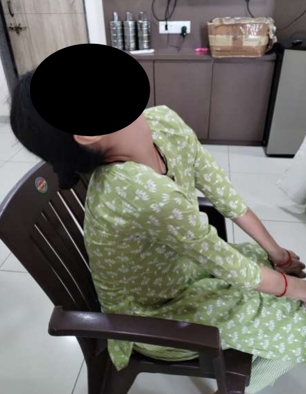
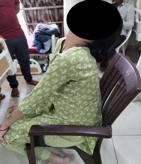
In this case, we have an antenatal patient of 24 years who experienced sudden jerky and continuous involuntary movements in the cervical spine during the beginning of her fourth month when she first visited us in Advance Hospital, Ahmedabad. Despite these movements, the patient remained conscious and oriented throughout, and her vital signs were stable. She complained the stiffness in neck, no numbness, slowness of ADL, imbalance while walking. It is worth noting that her antenatal course up until this point had been uneventful.
Medical History:
The patient's medical history includes a normal MRI brain with spine, normal ANA by IF, TSH, B2 glycoprotein, serum copper, ceruloplasmin, and normal ASO titre. Patient had no significant Past medical history.
Examination and Diagnosis:
Upon further examination, the patient was diagnosed with RHD (Rheumatic Heart Disease) upon undergoing 2d echo. This diagnosis sheds light on a potential underlying cause for the involuntary movements she experienced. While RHD can present with various symptoms, it is important to note that each case is unique and requires proper medical attention.
The patient was prescribed Trihexyphenidyl 2mg Benzhexol ½ for 5 days, clonazepam 0.5mg for 5 days, and tetrabenazine 25mg for 1 week without any prior thyroid disease history by the neurophysician alongwith her regular antenatal medicines prescribed by us.
The patient underwent planned cesserean section under General Anaesthesia and course of treatment and hospital stay was normal.
The fact that the patient consistently attended her prenatal visits on time demonstrates her commitment to maintaining a healthy pregnancy. It also highlights the importance of regular check-ups in identifying any potential complications or health issues.
Discussion and Conclusion
Overall, this case emphasizes the significance of comprehensive antenatal care and prompt diagnosis in ensuring both maternal and fetal well-being. By closely monitoring patients during their pregnancy journey, healthcare professionals can provide appropriate interventions and support to manage any unexpected developments effectively