11 Years Old Boy Presented with Unexplained Right Flank Mass and Negative Laboratory Results and Incidentally found to be Having Severe Left Hydrouretronephrosis and Vesicureteric Reflux.
11 Years Old Boy Presented with Unexplained Right Flank Mass and Negative Laboratory Results and Incidentally found to be Having Severe Left Hydrouretronephrosis and Vesicureteric Reflux.
Dr. Ahmed Moosa Yahya Assery *
*Correspondence to: Dr. Ahmed Moosa Yahya Assery, Paediatrics ambulatory care consultant. King Abdulaziz Medical City, National Guard Medical Affairs, Family Medicine-Ambulatory care, NGCSC.
Copyright
© 2015: Dr. Ahmed Moosa Yahya Assery. This is an open access article distributed under the Creative Commons Attribution License, which permits unrestricted use, distribution, and reproduction in any medium, provided the original work is properly cited.
Received: 12 December 2014
Published: 22 January 2015
Abstract
Having a strange-rare clinical diagnosis that linked with an eventual unsuspected sequala is something interesting, and this is what we have here in such case report for this 11 years old boy who came from the desert of KSA with a presentation taken subjectively from the father and documented later by a picture taken by the father mobile which was an appearing-disappearing left flank mass which was coming without clear other symptoms what obliged the father to do a blood and urine laboratory test in their nearest village which all came normal. Me in my clinic in addition to what was impressed about that mass (came later as LAWH) I discovered an (maybe) an incidental severs left HUNSIS and VUR on same side of the mass which needed an urgent RDSIS and further FU. Most of us aware about the challenge of discovering the VUR in children being most of the cases came asymptomatic but what interesting here is the left flank mass, its course of appearance and disappearance, is it related to the VUR we discovered, and any explanation of that association?
Keywords: KSA: Kingdom of Saudi Arabia.
HUNSIS: Hydroureteronephrosis.
VUR: Vesicoureteral Reflux.
RDSIS: Renal dialysis.
LAWH: Lateral Abdominal Wall Hernia.
FU: Follow Up.
UPJO: Ureteropelvic Junction Obstruction.
VCUG: Voiding Cystourethrogram.
UTI/s: urinary tract infection/s.
UVJ: ureterovesical junction.
11 Years Old Boy Presented with Unexplained Right Flank Mass and Negative Laboratory Results and Incidentally found to be Having Severe Left Hydrouretronephrosis and Vesicureteric Reflux.
Introduction
LAWH: The first description of a lateral abdominal hernia can be traced back to a case reported in 1906. While these defects occur less frequently than ventral abdominal hernias, they can progress to a very large size and cause significant morbidity.
Lateral abdominal wall hernias are a distinct entity from ventral abdominal wall hernias with respect to their anatomy, clinical behaviour, and surgical management. The ethology of the defect, presence of substantial static anatomic supporting structures, and extent of reinforcement have a significant impact on long-term recurrence rates. (1).
LAWH must be differentiated from other rare types of hernias such as lumbar hernia or spigelian hernia. There are various types of abdominal wall hernias. Common types of hernias such as inguinal hernia, femoral hernia, and umbilical hernia can be diagnosed easily by their locations. However, LAWH is rare. Differential diagnoses of this condition involve rare types of hernias such as lumbar hernia or spigelian hernia. Incisional hernias or traumatic hernias can also be developed, though rarely, in the lateral abdominal wall. Nonincisional or nontraumatic LAWH is an extremely rare condition. Spigelian hernia constitutes only 0.12% of all abdominal wall hernias. It is also known as 'spontaneous lateral ventral hernia' or 'hernia of semilunar line'. Spigelian hernia is a defect in the anterior abdominal wall beside the semilunar line. Most of these hernias lie in the spigelian hernia belt, a transverse 6-cm-wide zone above the interspinal plane. The present case is not located on the Spigelian hernia belt.
Lumbar hernias are rare posterolateral abdominal wall hernias. Primary lumbar hernias are classified into two types according to the location: the superior lumbar hernia (Grynfeltt) and the inferior lumbar hernia (Petit). They develop through the superior and inferior lumbar triangles. The superior lumbar triangle is bordered by the quadratus lumborum muscle medially, the posterior border of the internal oblique muscle inferiorly, and the 12th rib superiorly. The inferior lumbar triangle is bordered by the iliac crest inferiorly, the latissimus dorsi medially, and the external oblique muscle laterally. In the aspect of location, the present case is different from typical primary lumbar hernias. (2).
HUNSIS: (dilation of the renal pelvis) is a common, readily diagnosed finding on antenatal ultrasound examination, which may be a transient benign condition or be associated with significant congenital anomalies of the kidney and urinary tract. The goal of postnatal management of infants with prenatally diagnosed hydronephrosis is to identify those with clinically significant congenital anomalies of the kidney and urinary tract while avoiding unnecessary testing in patients with physiologic or clinically insignificant hydronephrosis. (3).
HUNSIS in the neonatal period can have a range of abnormalities including UPJO, ureterovesical junction obstruction, megacystis megaureter, or VUR. The second and third entities are uncommon and, therefore, paediatric patients who have hydronephrosis and a normal VCUG are presumed to have UPJO. This abnormality occurs in approximately 1 in every 2,000 live births and accounts for approximately half of the cases of prenatal hydronephrosis.
In most cases, neonatal hydronephrosis and presumed UPJO gradually resolves without surgical intervention. There is a strong correlation between the Society for Fetal Urology grade of hydronephrosis and the likelihood of spontaneous resolution: Grade I resolves in approximately 50% of patients, and grades II, III, IV hydronephrosis resolve in 36%, 16%, and 3% of cases, respectively.2 However, there is little information about the natural history of hydronephrosis and the time to resolution of the lesion in current practice when most women undergo serial sonography and screening for renal anomalies during pregnancy. Moreover, there are few guidelines regarding frequency of follow-up visits and referral to urology for more extensive testing such as performance of renal radionuclide scans and possible surgery in children with hydronephrosis who are cared for by general paediatricians and paediatric nephrologists. Additionally, the value of clinical markers such as birth history, UTI, and severity of the hydronephrosis as guides in predicting which patients are likely to require urological evaluation has not been adequately studied. Therefore, this review was conducted to determine the time course until spontaneous resolution of neonatal hydronephrosis and presumed UPJO and to identify risk factors for persistent hydronephrosis in a group of children followed by a paediatric nephrology service. The primary target audience of this review is general paediatricians to help guide strategies for serial observation and referral to paediatric urology. (4).
VUR: UR is the retrograde passage of urine from the bladder into the upper urinary tract. It is divided into two categories: primary and secondary based on the underlying pathogenesis.
Primary VUR — Primary VUR, the most common form of reflux, is due to incompetent or inadequate closure of the UVJ, which contains a segment of the ureter within the bladder wall (intravesical ureter). Normally, reflux is prevented during bladder contraction by fully compressing the intravesical ureter and sealing it off with the surrounding bladder muscles.
In primary VUR, failure of this anti-reflux mechanism is due to a congenitally short intravesical ureter. The intravesical ureter length may be genetically determined, which may explain the increased incidence in family members of patients with VUR.
Spontaneous resolution of primary low-grade VUR commonly occurs with patient growth. As the bladder grows, the intravesical ureter increases in length, improving the function of the anti-reflux mechanism.
Secondary VUR — Secondary VUR is a result of abnormally high voiding pressure in the bladder that results in failure of the closure of the UVJ during bladder contraction. Secondary VUR is often associated with anatomic (eg, posterior urethral valves) or functional bladder obstruction (eg, bladder bowel dysfunction [BBD] and neurogenic bladder). The degree and chronicity of obstruction can influence the severity of VUR. (5).
Our Case Presentation
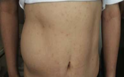
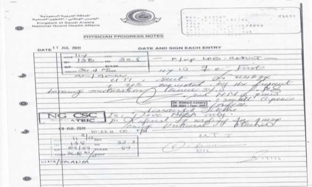
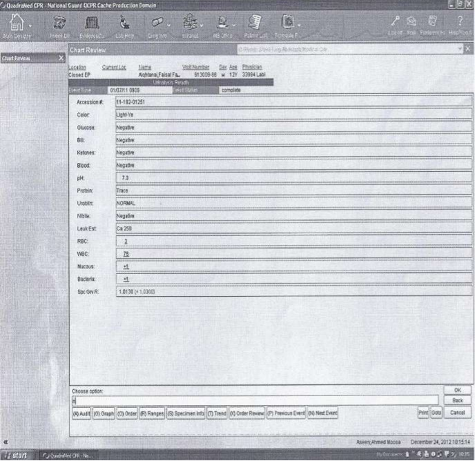
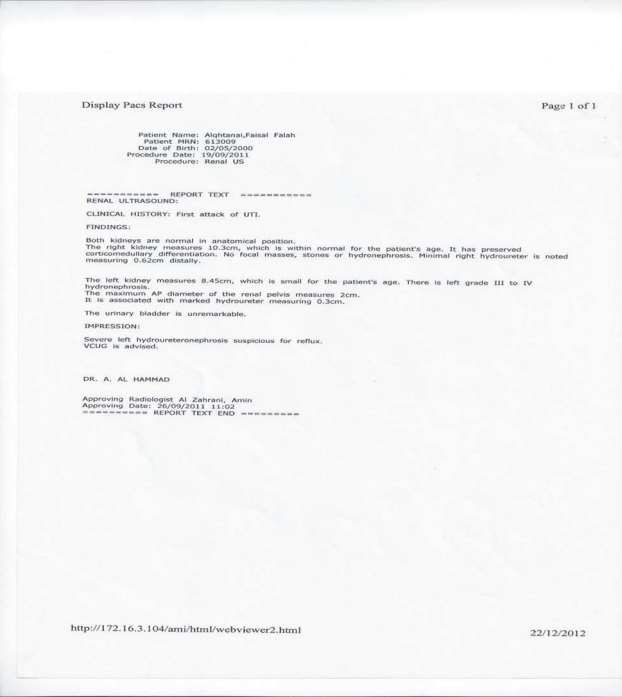
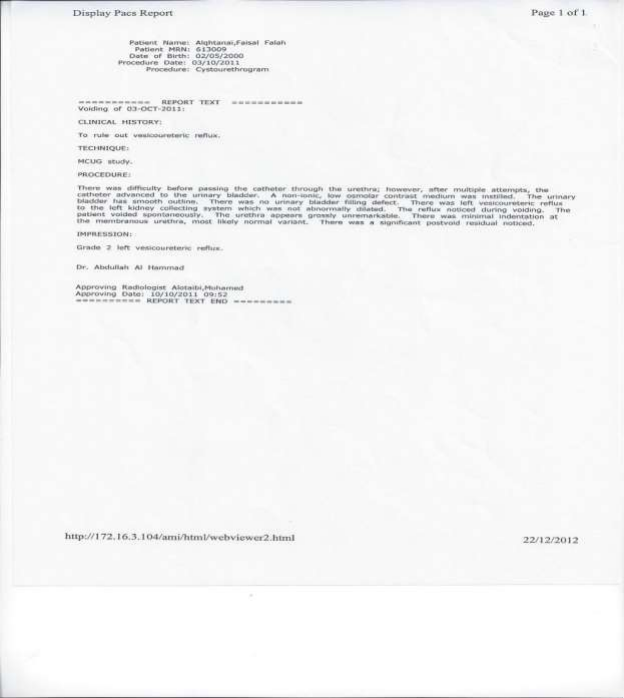
This child who was 11 years old boy came ( with his father ) from the desert of KSA, an area that almost 300 kilometre far from the capital concerning about a swelling on the left flank, first appeared when he was 9 years old , not preceded by any trauma or surgeries, that swelling not associated with any symptoms at all except what was strange swelling was appearing and disappearing alternatively which made the father take pictures of that sweeling showed in ( Figure No 1): and since of the extreme poverty that family living in they visited anear by primary health centre that done for the child simple laboratory blood test and advised the family to seek a medical advice in more qualified medical centre having a surgery team to investigate further about this abdominal mass. The patient booked with me on 11 July 2011 (Figure No 2): and among the routine investigations requested the initial urine analysis (Figure No 3): that came suggestive partially of UTI despite the negative nitrite despite the negative urine culture (Figure No 4) after which I decided to offer for the child a renal ultrasound at least before the paediatrics surgery consultation regarding to the abdominal mass. The surprise came which was the radiology that was suggestive of sever left HUNSIS ranging between grades III and IV (Figure No 5) that why the I consulted the pediatrics urology who preceded with VCUG in the next available chance. The VCUG confirmed the presence of the left HUNSIS with presence of grade 2 VUR on the same side(Figure No 6).
The patine soon undergone RDSIS and among the admission seen by the paediatric surgery since of the right abdominal mass and found to be a LAWH that should be repaired soon after solving the problem of the VUR.
Figure No 1: Our case presentation flank mass on the right side of The abdomen that was labeled as LAWH.
Figure No 2: The progress note of the patient visit to my clinic.
Figure No 3: The initial urine analysis that came with high white blood cells of 78/hfp
Figure No 4: The urine culture came mostly with picture of contaminated sample.
Figure No 5: The radiology report of the ultrasound of the patient that came suggestive of left HUNSIS.
Figure No 6: The VUCG report.
Discussion about the Case
So interesting that me and the family were concerned about the presenting complaint (the abdominal mass) and later we discovered this disaster we cough it early before the complete damage of the left kidney. To correlate the patients LAWH and VUR this needs deep search in the Medline medical articles which never ever linked them in any place. Because the VUR is relatively common in pediatrics with a prevalence for children with febrile urinary tract infections (UTIs) (ranging from 30 to 45 percent) (5) it is fair here to review the LAWH case reports because it is very rare and search its relations if found with other body systems defects like that one in our case.
In national library of medicine journal Ann Med Surg (Lond). 2023 Apr; 85(4): 1194–1196.Published online 2023 Apr 6. doi: 10.1097/MS9.0000000000000454 published by Mabrouk Abdelali, MD,a Mohamed Ali Chaouch, MD, b Sadok Ben Jabra, MD,b Jamal Saad, MD,a Maha Ben Mansour, MD,c Sawsen Chakroun, MD,c Yasmine Khouni, MD,a Fadwa Aguir, MD,a Asma Achour, MD,a Ahmed Zrig, MD,a Faouzi Noomane, MD,b and Mezri Maatouk, MDa under the title Post-traumatic lateral abdominal wall hernia: a case report, Concluded that TAWH should be suspected behind any blunt abdominal trauma with high energy. CT scan and ultrasound were helpful for diagnosis and the only curative treatment is surgery to avoid complications. (6).
In Indian Journal of Radiology and Imaging Indian J Radiol Imaging 2024; 34:377–379. Published by Siddhi Chawla1 Aditya Charan1 under the title: concluded that Experience with congenital LAWH is limited, with only 60 cases reported in the literature.3,4 The probable cause for development of LAWH includes insult during embryonic development causing defect in the germinal disk, defects in the amnion, defects in the ectodermal placode, and/or vascular defects. Nonvisualization of all the abdominal wall layers with maintained skin places the time of insult probably during embryogenesis. (7).
In Plastic and reconstructive surgery journal 11(9): p e5007, September 2023. | DOI: 10.1097/GOX.0000000000005007 published by Mattison, Gennaya Lynn MD*; Lee, Seung Ah MD*; Wong, Daniel Enjay MD†; Leevan, Elyse MD‡; Carmichael, Joseph Christopher MD‡; Evans, Gregory Randolph Dean MD* under the title Recurrent Lateral Abdominal Wall Hernias: Options for Reconstruction – case report mentioned that This case details a recurrent flank hernia originally caused by blunt abdominal trauma, repaired by an underlay mesh with anchoring sutures, transfascial sutures, and tacks. These injuries remain rare, with limited reports in the literature. Additional clinical data and review continue to contribute to our knowledge and improved management of these complex hernias. (8).
As you saw from the reviews above that LAWH is extremely rare and mostly came as a sequalae of a sever blunt trauma of the abdomen and no relation at all with any adjacent body systems including the urinary one.
Conclusion
The LAWH still an extremely rare surgical disorder that most of them are post-traumatic except our case here that was not but could VUR be one of the causative factors? We need further case reports and researches to confirm or deny.
References
1-https://www.uptodate.com/contents/lateral-abdominal-wall-hernia-repair?search=lateral%20abdomenal%20wall%20hernia&source=search_result&selectedTitle=1%7E8&usage_type=default&display_rank=1.
2-https://www.ncbi.nlm.nih.gov/pmc/articles/PMC4325649/.
3-https://www.uptodate.com/contents/postnatal-evaluation-and-management-of-hydronephrosis?search=Hydroureteronephrosis.&source=search_result&selectedTitle=2%7E7&usage_type=default&display_rank=2.
4-https://www.ncbi.nlm.nih.gov/pmc/articles/PMC3676294/.
5- https://www.uptodate.com/contents/clinical-presentation-diagnosis-and-course-of-primary-vesicoureteral-reflux?search=vesicoureteral%20reflux&source=search_result&selectedTitle=1%7E129&usage_type=default&display_rank=1.
6-https://www.ncbi.nlm.nih.gov/pmc/articles/PMC10129220/.
7-https://www.thieme-connect.com/products/ejournals/pdf/10.1055/s-0043-1777290.pdf.
8-https://journals.lww.com/prsgo/fulltext/2023/09000/recurrent_lateral_abdominal_wall_hernias__options.60.aspx.