Use of Oral Methotrexate and Misoprostol in the Treatment of Cesarean Scar Pregnancy
Use of Oral Methotrexate and Misoprostol in the Treatment of Cesarean Scar Pregnancy
Khadeeja Yahya Osman *1, Roaa Mohamed Ahmed Elhaj 2
1. Khadeeja Yahya Osman, OB/GYN Specialist, King Fahad Hospital, MBBS( University of Medical Science and Technology), Clinical MD in Obstetrics and Gynecology (Sudan Medical Specialization Board), MSC in advanced Ultrasound in Obstetrics and Gynecology, MRCOG.
2.Roaa Mohamed Ahmed Elhaj, OB/GYN Specialist, Shaqra General Hospital, MBBS (University of Khartoum ), Clinical MD in Obstetrics and Gynecology (Sudan Medical Specialization Board), MRCOG, MSC Medical Education (Warwick University).
*Correspondence to: Khadeeja Yahya Osman, ORCID: 0000-0002-5725-9350.
Copyright
© 2024 Khadeeja Yahya Osman. This is an open access article distributed under the Creative Commons Attribution License, which permits unrestricted use, distribution, and reproduction in any medium, provided the original work is properly cited.
Received: 12 August 2024
Published: 21 August 2024
Use of Oral Methotrexate and Misoprostol in the Treatment of Cesarean Scar Pregnancy
Introduction
Cesarean scar pregnancy (CSP) is defined as a pregnancy in a previous cesarean scar. It has high risks for severe maternal morbidities like uterine rupture, hemorrhage, or live births complicated by placenta accreta spectrum (PAS), hemorrhage, and/or risk of cesarean hysterectomy [1]. In addition, it’s likely that patients with a prior CSP are at higher risk of recurrence [2]. Published rates for the recurrence of CSP have been estimated between 17.6% and 34.3% [3].
Cesarean scar pregnancy (CSP) is rare. Estimated range varies from 1:1,800 to 1:2,656 of overall pregnancies since 2000s, however ,the incidence of CSP is increasing due to the increasing rate of cesarean section deliveries [7]. A previous study from 2011 based at a big tertiary care hospital in Israel estimated the incidence of CSP as 1 per 3,000 for the general obstetric population and 1per 531 among the patients who had at least one cesarean delivery [8]. The same study also stated that, CSP constituted 4.2% of ectopic pregnancies [9]. When the patient is diagnosed to have CSP, observative management is not recommended by the Society of Maternal-Fetal Medicine (SMFM) [10]. Several treatments and a combination of treatments for CSP have been reported in the literature, however, the optimal treatment is still unclear [11]. the SMFM Consult series, recommendations for treatment of CSP pregnancies include active management with operative resection, ultrasound-guided suction dilation and curettage (D&C), or intra-gestational methotrexate with or without other modalities. [12]. Mifepristone, is usually used in combination with misoprostol for abortion management. It is another drug that may be used in CSP jointly with other methods, although it not described in the SMFM Consult series [13].
Objective
To evaluate the effective ness of the use of Oral Methotrexate and misoprestole in the treatment of cesarean scar pregnancy.
Case Presentation
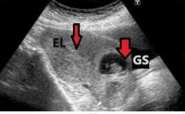
The Patient was 39-years-old G5P4 all previous deliveries were by with cesarean sections last one 2 years ago. The patient presented with amenorrhea and was found to have a positive urine pregnancy test. It was an unplanned pregnancy, but desired. At 7 weeks gestational age, she had her first transvaginal ultrasound, which showed a pregnancy concordant with her last menstrual period with cardiac activity. there was a concern for implantation in her cesarean scar indicating cesarean scar pregnancy (SCP). see pic 1.
Pic 1: TVS shows: clear endometrial line and csp
She was counseled about CSP and associated risks. It was recommended to terminate the pregnancy. Treatment options included operative management with suction D&C, cervical ripening balloon placement in addition to gravid hysterectomy. Medical treatment was also offered to be used with any of the operative options. She desired to repeat the scan for verification of CSP as this pregnancy was highly desired if not a CSP. Scan repeated at 8 weeks. With her repeat ultrasound at 8 weeks gestation, she was again noted to have a pregnancy implanted in her previous cesarean scar and had trophoblastic tissue extending to within 1.5 millimeters (mm) of the anterior uterine wall, without evidence of invasion into bladder. Again, this was discussed with the patient, and she wished to proceed with treatment at that time. Although she did not plan future fertility, she strongly desired to avoid a hysterectomy.
She was recommended medical and surgical management jointly, depending on availability of operating room, desire for cervical preparation, and preference to end fetal cardiac activity prior to the procedure if possible.
Systemic methotrexate was not available, so she had to choose between referral to higher center or to start oral methotrexate. At the end, she received oral methotrexate with 60 mg/m2 , as. see follow up chart 1.
|
|
Day 0 |
Day 4 |
Day 7 |
|
B HCG |
4500 |
5100 |
4670 |
|
Renal function test |
Normal |
|
Normal |
|
Liver function test |
Normal |
|
Normal |
|
Vital signs |
Normal |
Normal |
Normal |
|
Concerning symptoms |
-ve |
-ve |
-ve |
|
TVS |
Viable, scar pregnancy, CRL 8wks +4days |
|
+VE FH |
Reassessment was done including TVS which revealed still positive fetal heart. Bhcg was dropping but less that 15%, pt was vitally stable, renal and liver functions were normal , after proper counselling an another dose of methotrexate of 60 mg/m2 was given. and she had been followed on day 0,4,7 see follow up chart 2.
|
|
Day 0 |
Day 4 |
Day 7 |
|
B HCG |
4670 |
4325 |
3340 |
|
Renal function test |
Normal |
|
Normal |
|
Liver function test |
Normal |
|
Normal |
|
Vital signs |
Normal |
Normal |
Normal |
|
Concerning symptoms |
-ve |
-ve |
-ve |
|
TVS |
+ve fetal heart |
|
-ve fetal heart |
Chart 2: Follow up after second Methotrexate dose
She was planned for weekly follow up of beta hcg , one week later she presented to ER complaining of abdominal cramping and vaginal bleeding . she was vitally stable TVS revealed incomplete miscarriage . she received sublingual misoprostol 600 mic. Later during the day she developed sever pain and expelled products adequate analgesia and rehydration fluid were offered to stabilize the patient . she expelled products after which she received one dose of intramuscular methergine due to brisk of vaginal bleeding after which she received a 24-hour course of oral methergine. She was monitored in the hospital overnight with minimal bleeding, and she was discharged 1 day later after confirming complete miscarriage evidence by TVS. See pic 1.
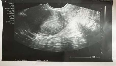
Three weeks later urine pregnancy test was negative. There were no complications. Histopathology confirmed products of conception. Counselled about contraception, she was offered subdermal implants.
Pic 2: TVS Shows: Bulky anteverted uterus, empty with clear endometrial line indicates complete miscarriage
Discussion
Methotrexate is a chemotherapeutic agent, acting as an anti-neoplastic anti metabolite with immunosuppressant activity . Its usage in gynecology was first reported by Li and Heartz in 1956 where it was used to treat patients with choriocarcinoma. methotrexate works by blocking the DNA synthesis as it works by blocking the folic acid binding site on the enzyme dihydrofolate reductase thus rendering it inactive hence prevent conversion of folic acid to tetrahydro folic acid . Tetrahydro folic acid is important in the DNA synthesis process as it suport the one-carbon transfer reaction needed by the coenzymes. [14].
This was an example of CSP, which was successfully treated with oral methotrexate and misoprostol without need for surgical interventions. Although most of studies confirmed the role of systemic methotrexate in the treatment of CSP, there are some studies supporting the use of oral methotrexate. There is a cross-sectional study using secondary data obtained from medical record office on patients with ectopic pregnancy included a total of 35 patients who fulfilled the criteria for medical management.
They were prescribed with oral methotrexate with the dose of 60 mg given in 3 divided doses every 2 hours using the standard tablet of 2.5mg. Follow up was done at day 4, day 7 and till HCG level achieve less than 20 iu/litre, success rate was 82% without need for other intervention. The side effects of oral methotrexate were tolerated well by all patients which supports the condition of the current case report.
The advantages of using lower dose of methotrexate includes the reduction in side effects of methotrexate and folic acid rescue was not needed. Another advantage of oral methotrexate over intramuscular methotrexate is that the administration via intramuscular route trigers pain and local reaction to surrounding soft tissues.
Furthermore, Oral methotrexate is more cost effective as we need less equipment and expertise (pharmacist in handling parenteral cytotoxic drug).
Conclusion
In conclusion, the inaccessibility to parenteral methotrexate has opened a new horizon to find out other options which will benefit more women in the future. Oral methotrexate can be used successfully to treat ectopic pregnancy and there are few advantages over intramuscular methotrexate. This Case reports , opens the way forward for more work up on the use of oral methotrexate and development of more guide lines to support providing good clinical treatment with minimal facilities available and less side effects.
References
1.Cesarean scar pregnancy: issues in management. Seow KM, Huang LW, Lin YH, Lin MY, Tsai YL, Hwang JL. Ultrasound Obstet Gynecol. 2004;23:247–253.
2.Fertility performance and obstetric outcomes among women with previous cesarean scar pregnancy. Maymon R, Svirsky R, Smorgick N, Mendlovic S, Halperin R, Gilad K, Tovbin J. J Ultrasound Med. 2011;30:1179–1184.
3.Cesarean scar ectopic pregnancy: imaging features, current treatment options, and clinical outcomes. Riaz RM, Williams TR, Craig BM, Myers DT. Abdom Imaging. 2015;40:2589–2599.
4.Society for Maternal-Fetal Medicine (SMFM) Consult Series #49: cesarean scar pregnancy. Miller R, Timor-Tritsch IE, Gyamfi-Bannerman C. Am J Obstet Gynecol. 2020;222:0.
5.First-trimester diagnosis and management of pregnancies implanted into the lower uterine segment Cesarean section scar. Jurkovic D, Hillaby K, Woelfer B, Lawrence A, Salim R, Elson CJ. Ultrasound Obstet Gynecol. 2003;21:220–227 .
6. Ectopic pregnancies in a Caesarean scar: review of the medical approach to an iatrogenic complication. Maymon R, Halperin R, Mendlovic S, Schneider D, Herman A. Hum Reprod Update. 2004;10:515–523.
7.Caesarean scar pregnancy. Ash A, Smith A, Maxwell D. BJOG. 2007;114:253–263.
8.Caesarean scar pregnancy. Ash A, Smith A, Maxwell D. BJOG. 2007;114:253–263.
9.Reproductive outcome after cesarean scar pregnancy: a systematic review and meta-analysis. Morlando M, Buca D, Timor-Tritsch I, et al. Acta Obstet Gynecol Scand. 2020;99:1278–1289.
10.Jabarudin WNEW et al. Int J Reprod Contracept Obstet Gynecol. 2019 Aug;8(8):3229-3233
11.First-trimester diagnosis and management of pregnancies implanted into the lower uterine segment Cesarean section scar. Jurkovic D, Hillaby K, Woelfer B, Lawrence A, Salim R, Elson
12.CJ. Ultrasound Obstet Gynecol. 2003;21:220–227 .
13.Society for Maternal-Fetal Medicine (SMFM) Consult Series #49: cesarean scar pregnancy. Miller R, Timor-Tritsch IE, Gyamfi-Bannerman C. Am J Obstet Gynecol. 2020;222:0.
14.Jabarudin WNEW et al. Int J Reprod Contracept Obstet Gynecol. 2019 Aug;8(8):3229-3233