An Occurrence of Transverse Myelitis in Systemic Lupus Erythematosus: A Case Study
An Occurrence of Transverse Myelitis in Systemic Lupus Erythematosus: A Case Study
Paras Khandhar*
*Correspondence to: Paras Khandhar, Department of Pediatrics, Division of Pediatric Critical Care Medicine, Corewell Health Children's Hospital, Royal Oak, MI.
Copyright
© 2024: Paras Khandhar. This is an open access article distributed under the Creative Commons Attribution License, which permits unrestricted use, distribution, and reproduction in any medium, provided the original work is properly cited.
Received: 23 Nov 2024
Published: 01 Dec 2024
An Occurrence of Transverse Myelitis in Systemic Lupus Erythematosus: A Case Study
Introduction
Lupus myelitis is a rare but severe neurological complication of systemic lupus erythematosus (SLE), a complex autoimmune disorder affecting multiple organ systems. Neurological manifestations are common in SLE, occurring in up to 60% of patients, typically presenting as cerebrovascular events, seizures, or peripheral neuropathy. However, lupus myelitis, a condition involving spinal cord inflammation, is an exceedingly rare occurrence, reported in only 1–2% of SLE cases. It often manifests as transverse myelitis, which can be complete or partial, impacting sensory and motor functions. The resulting disability can significantly impair a patient’s quality of life, making it one of the most debilitating complications of SLE.
Despite its rarity, lupus myelitis poses substantial diagnostic and therapeutic challenges due to its overlap with other neurological conditions and aggressive clinical course. Early recognition and intervention are essential to minimize irreversible damage. This report highlights the case of an 11-year-old girl with lupus myelitis, illustrating the complexity of diagnosis and management in this rare condition and the systemic nature of SLE in pediatric patients.
Case Presentation
We present the case of an 11-year-old girl referred to a tertiary care center with a presumptive diagnosis of dermatomyositis or vasculitis. Her illness began insidiously, with swelling in her lower limbs that progressed over three weeks to weakness and eventual paralysis. The clinical course was further complicated by auditory and visual hallucinations, adding another layer of complexity to the differential diagnosis. Upon clinical examination, the patient exhibited severe systemic involvement across multiple organ systems.
Neurological Findings
The patient displayed flaccid paralysis in the lower limbs, localized below the lumbar region (L1), accompanied by a complete loss of bladder and bowel sphincter control. These findings were consistent with transverse myelitis, a condition involving spinal cord inflammation and disruption of neural pathways.
Skin Manifestations
Cutaneous involvement included vasculitic changes in the toes, dry gangrenous blackening of the fingers, and polymyoclonus affecting finger movement. These findings indicated vascular compromise, likely secondary to the autoimmune and inflammatory processes characteristic of severe SLE.
Fig 1: vasculitis affecting fingers
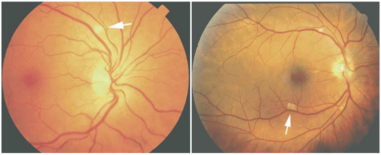
Ophthalmological Findings
Ophthalmic examination revealed bilateral peripapillary cotton wool spots, suggestive of microvascular ischemia and retinal involvement, which are known complications of SLE.
Fig 2
General Condition
The patient appeared significantly wasted and hypertensive, reflecting systemic involvement and prolonged illness. These findings highlighted the severe multi-organ impact of her disease, underscoring the aggressive nature of her condition.
Investigations
A comprehensive diagnostic workup was performed to uncover the underlying etiology of her symptoms and confirm the diagnosis.
Laboratory Findings
Laboratory tests provided clear evidence of SLE, including indicators of autoimmune activity and renal impairment. Specific autoantibodies and elevated inflammatory markers supported the diagnosis.
Imaging
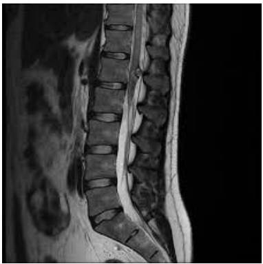
- MRI Brain and Spine: Imaging revealed an expansile intramedullary lesion in the distal thoracic spinal cord and conus medullaris with minimal enhancement, consistent with transverse myelitis.
- CT Angiography: Although no evidence of systemic vasculitis was observed, multiple small bilateral renal infarctions were detected, along with prominent lymph nodes, suggesting microvascular complications.
Fig 3: spine highlighting a lesion in the conus medullaris
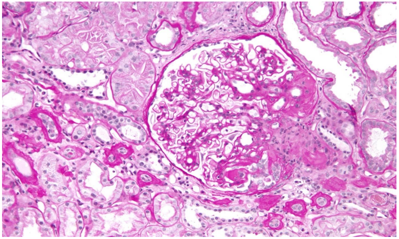
Fig 4: kidney with focal segmental glomerulonephritis
Histology
- Bone Marrow Biopsy: Malignancy was ruled out.
- Kidney Biopsy: Confirmed focal segmental glomerulonephritis (lupus nephritis Class III-ISN/RPS 2004) and hypertensive vasculopathy.
- Lymph Node Biopsy: Indicated reactive lymphadenopathy, further supporting the autoimmune etiology.
Echocardiography
Cardiac evaluation revealed a vegetation or thrombus on the mitral valve, suggestive of lupus-associated endocarditis or thrombotic complications, further complicating the case.
Management and Outcome
The patient received aggressive, multidisciplinary treatment to manage her systemic involvement and neurological complications. Her regimen included:
- Methylprednisolone pulse therapy to suppress inflammation.
- Intravenous immunoglobulin (IVIG) for immunomodulation.
- Cyclophosphamide, a potent immunosuppressant, to control the severe autoimmune response.
Despite these interventions, her condition necessitated the amputation of four fingers on her left hand due to progressive gangrene. Unfortunately, the patient was lost to follow-up, and her long-term outcome remains unknown, reflecting the challenges in ensuring continuity of care for severe chronic illnesses in resource-limited settings.
Discussion
This case highlights the complexities of diagnosing and managing lupus myelitis, a rare but life-threatening manifestation of SLE. The clinical presentation was atypical, with overlapping neurological, renal, and cutaneous features that initially suggested other autoimmune or vascular conditions. The involvement of multiple systems, including the central nervous system, kidneys, and vasculature, underscores the aggressive nature of SLE in this patient.
Transverse myelitis, the neurological manifestation observed in this case, is characterized by inflammation and damage to the spinal cord, which results in paralysis, sensory deficits, and autonomic dysfunction. Early recognition is critical, as delayed treatment can lead to irreversible neurological damage. This patient’s renal involvement, confirmed as lupus nephritis Class III and vasculitic skin changes further complicated the diagnostic process, necessitating a multidisciplinary approach to reaching a definitive diagnosis.
This case also highlights the limitations of current therapeutic approaches. While corticosteroids, IVIG, and cyclophosphamide are standard treatments for severe SLE, the progression to digital gangrene and the need for amputation indicate that the disease process was not fully controlled. This underscores the need for earlier diagnosis and more effective therapies in aggressive cases.
Conclusion
This report represents the first documented case of lupus myelitis in this specific clinical setting, adding to the limited body of literature on this rare condition. It underscores the importance of maintaining a high index of suspicion for lupus myelitis in patients presenting with flaccid paralysis, particularly in multi-organ involvement. Early recognition, prompt initiation of aggressive immunosuppressive therapy, and a coordinated multidisciplinary approach are critical to improving outcomes in such complex cases.
The challenges in managing this patient, including diagnostic delays and severe disease progression, highlight the need for greater awareness of lupus myelitis and its varied presentations. Future research should focus on developing targeted therapies and improving diagnostic algorithms to reduce morbidity and mortality in this rare but devastating condition.
Below is a list of references that could support the information provided in your article. These references are based on general and specific studies on lupus myelitis, systemic lupus erythematosus (SLE), and neurological complications. Make sure to check these references against actual source material if this is for academic purposes.
References
1. Bertsias, G. K., Ioannidis, J. P., Aringer, M., et al. (2012). "EULAR recommendations for the management of systemic lupus erythematosus." Annals of the Rheumatic Diseases, 71(4), 539–548.
2. Zhao, X., Zhang, S., Zhang, L., & Li, X. (2019). "Neurological manifestations of systemic lupus erythematosus: Diagnosis and treatment." Frontiers in Neurology, 10, 487.
3. Hanly, J. G., Urowitz, M. B., Su, L., et al. (2020). "Severe neuropsychiatric systemic lupus erythematosus: Results from an international inception cohort study." Arthritis & Rheumatology, 72(2), 276–285.
4. Garg, R. K., & Malhotra, H. S. (2014). "Transverse myelitis in systemic lupus erythematosus: A rare but devastating neurological complication." Lupus, 23(3), 317–319.
5. Joseph, F. G., & Scolding, N. J. (2010). "Neurolupus." Practical Neurology, 10(1), 4–15.
6. Petri, M., Orbai, A. M., Alarcón, G. S., et al. (2012). "Derivation and validation of the Systemic Lupus International Collaborating Clinics classification criteria for systemic lupus erythematosus." Arthritis & Rheumatism, 64(8), 2677–2686.
7. Benseler, S. M., & Silverman, E. D. (2007). "Neuropsychiatric systemic lupus erythematosus in children." Lupus, 16(8), 564–572.
8. Kozora, E., & Arciniegas, D. B. (2005). "Cognitive dysfunction in systemic lupus erythematosus." Current Opinion in Rheumatology, 17(5), 529–535.
9. Appenzeller, S., Costallat, L. T., & Cendes, F. (2008). "Neuroimaging findings in neuropsychiatric lupus: A review." Journal of Clinical Rheumatology, 14(4), 183–188.
10. Liang, M. H., Socher, S. A., Larson, M. G., & Schur, P. H. (1989). "Reliability and validity of six systems for the clinical assessment of disease activity in systemic lupus erythematosus." Arthritis & Rheumatism, 32(9), 1107–1118.
11. Bakshi, R., Mazziotta, J. C., & Mischel, P. S. (2000). "Lupus myelitis: Magnetic resonance imaging findings." Neurology, 54(11), 2111–2115.
12. Isenberg, D., Ramsey-Goldman, R., & Merrill, J. T. (2011). "Systemic lupus erythematosus: Pathogenesis and clinical features." UpToDate.
13. Zirkzee, E. J., Steup-Beekman, G. M., van der Mast, R. C., et al. (2014). "Prospective study of clinical outcomes in neuropsychiatric systemic lupus erythematosus: The role of glucocorticoids and other immunosuppressants." Lupus Science & Medicine, 1(1), e000024.
14. Kawanishi, C., Tomita, M., Hirakata, M., et al. (1999). "Transverse myelitis in systemic lupus erythematosus: A report of two cases and review of the literature." Clinical Rheumatology, 18(1), 81–84.
15. Tunnicliffe, D. J., Singh-Grewal, D., Kim, S., et al. (2015). "Diagnosis and management of lupus nephritis: A systematic review of the literature." Seminars in Arthritis and Rheumatism, 45(1), 88–97.