An Update on PCOS Pathophysiology in Reference to Oxidative Stress (Os), Ferroptosis: A Comprehensive Narrative Review on Part of N-Acetylcysteine (Nac), & Selenium in Tackling Ferroptosis, Os & PCOS
An Update on PCOS Pathophysiology in Reference to Oxidative Stress (Os), Ferroptosis: A Comprehensive Narrative Review on Part of N-Acetylcysteine (Nac), & Selenium in Tackling Ferroptosis, Os & PCOS
Dr Kulvinder Kochar Kaur *1; Dr Gautam Nand Allahbadia 2, Dr Mandeep Singh 3
1) Dr Kulvinder Kochar Kaur, M.D (Obstt &Gynae, specialist reproductive endocrinology & Infertility specialist).
Scientific Director cum Owner Dr Kulvinder Kaur Centre For Human Reproduction, 721,G.T.B. Nagar, Jalandhar-144001, Punjab, India. Orcid Number- https://orcid.org/0000-0003-1473-3419.
2) Dr Gautam Nand Allahbadia M.D.(Obstt&Gynae),D.N.B, Scientific Director, Ex-Rotunda-A Centre for Human Reproduction 672, Kalpak Garden, Perry Cross Road, Near Otter’s Club,Bandra(W)-400040 Mumbai, India.
3) Dr Mandeep Singh M.D.DM.(Std)(Neurology), Consultant Neurologist, Swami Satyanand Hospital, Jalandhar-144001, Punjab, India
*Correspondence to: Dr Kulvinder Kochar Kaur, Scientific Director cum Owner Dr Kulvinder Kaur Centre For Human Reproduction, 721,G.T.B. Nagar, Jalandhar-144001, Punjab, India.
Copyright
© 2025 Dr Kulvinder Kochar Kaur. This is an open access article distributed under the Creative Commons Attribution License, which permits unrestricted use, distribution, and reproduction in any medium, provided the original work is properly cited.
Received: 21 April 2025
Published: 06 May 2025
DOI:https://doi.org/10.5281/zenodo.15387844
ABSTRACT:
Previously we reviewed articles on polycystic ovary syndrome (PCOS) pathophysiology, classification /therapy inclusive of insulin sensitizers like (D-chiro (DCI)- Myoinositol (M). N-acetylcysteine (NAC), a compound recognized for its cysteine and glutathione precursor characteristics, has been used as therapeutic applications for2 decades. Recently, an escalating attraction in evaluating the plausible advantages of NAC in tackling PCOS . Nonetheless, the precise mechanistic modes behind NAC’s therapeutic, and clinical utility continue to be uncharted. The objective of this narrative review was to investigate the manner NAC yields protection against PCOS and part of oxidative stress( OS) and oxidative reductive stress in the pathophysiology of PCOS and ferroptosis in PCOS. Therefore as a corollary Selenium, is a crucial constituent of glutathione peroxidase(GPx),an enzyme significant in ferroptosis and Thyroid hormone generation . Here we conducted a narrative review utilizing search engine pubmed,google scholar ;web of science ;embase; Cochrane review library utilizing the MeSH terms like polycystic ovary syndrome (PCOS); N-acetylcysteine (NAC); Selenium; oxidative stress( OS); ferroptosis from 2000 onwards till date in 2025. We found a total of 300 articles out of which we selected 71 articles for this review.No meta-analysis was done. Maximum studies point that NAC, if used alone or in combination with other medications, has the plausibility of countering OS, utilize its anti-inflammatory and anti-apoptotic characteristics, and yield advantages in PCOS management. Furthermore, NAC might possess the capability of impacting particular signaling pathways in insulin target cells and β cells. Variable biological actions of NAC suggest its plausible utility as a supplementary /therapeutic strategy for PCOS management. Therefore, further work is needed for evaluating plausible usefulness of NAC in tackling PCOS. Additionally, we detail how ferroptosis might be implicated in the pathophysiology of PCOS and Selenium is one more antioxidant of utility in PCOS therapy.
Key Words; polycystic ovary syndrome (PCOS); Nacetylcysteine (NAC); ); Selenium; oxidative stress( OS); ferroptosis.
An Update on PCOS Pathophysiology in Reference to Oxidative Stress (Os), Ferroptosis: A Comprehensive Narrative Review on Part of N-Acetylcysteine (Nac), & Selenium in Tackling Ferroptosis, Os & PCOS
Introduction
Polycystic ovary syndrome (PCOS) represents a substantially frequent endocrine condition, which influences women in developed as well as developing countries, with prevalence of variations differening from 8-13%[1,2]. Clinical diagnosis of PCOS gets made with the utilization of Rotterdam’s criteria [3] (any two of the following three criteria after similar exclusion criteria, i)clinical(for instance acne, hirsutismH ) in addition to /or biochemical androgen excess,ii) chronic anovulation situations (oligoovulation or anovulation till amenorrhea,O) along with iii)micropolycystic morphology on ultrasonology transducer having a frequency bandwidth 8MHZ (≥20 follicles/ ovary,or an ovarian volume ≥10ml on any ovaryP. Classification of PCOS is done into 4 subkinds dependent on criteria- TypeA( H+ O+P) Type B( H+ O ), Type C( H+ O+P) TypeD ( O+P) Such organization of PCOS differ as per racial as well as ethnic factors in addition to that once groups shift from one region to other regions,they have sustenance of susceptibility of their ethnicity to the generation of the PCOS along with the dysfunctional metabolism whose sustenance takes place secondary to hyperinsulinemia as well as/or diabetes mellitus(DM) as found for instance in women now based in Britain but are basically from Indian subcontinent Asian origin, who have greater prevalence of PCOS in addition to T2DM[4]. Such patients of PCOS frequently have associated metabolic as well as reproductive abnormalities iinclusive of obesity, insulin resistance (IR), oxidative stress(OS), acne, hirsutism,irregular periods, infertility, lesser pregnancy outcomes. Additionally, they have pronounced long term escalated complications for instance cardiovascular disease (CVD) as well as cancer[3] .
Uptill now the PCOS etiology continues to be uncharted . Despite variable hypothesis have been posited in reference to exposition of their pathogenic mechanistic mode of PCOS, for instance IR ,OS ,hyperinsulinemia as well as hyperandrogenism possess a crucial part[5,6]. Additionally, studies have pointed that OS is correlated with IR, hyperinsulinemia, hyperandrogenism, chronic inflammatory states, abnormalities of glucose in addition to lipid metabolism ,ovarian impairment, along with inimical pregnancy outcomes [7-9]. Hence, antioxidant therapies might be of help in the treatment of PCOS.
N-Acetyl Cysteine (NAC) represents a robust synthetic anti oxidant that forages superoxide,H2O2 , besides hydroxyl radicals in addition to restoration of glutathione(GSH) quantities, which might work in the form of a coenzyme in variable reactions .In its reduced state, it has an active sulf hydryl group that possesses the capacity of generation of disulfide bonds with free S-H groups rivalling · or deconstructing the existent disulfidebonds, working in the form of a reducing agent[10]. NAC has displayed a plethora of biological actions for instance anti apoptotic[11], working in the form of an anti oxidant system [12], resulting in avoidance of localized ischaemia[13], as well as blockade of for instance phospholipid(PL), metabolism, therefore liberation of proinflammatory cytokines in addition to protease actions[14].
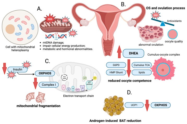
Effects of NAC are substantially complex in case of PCOS[15-17]. Is NAC capable of treatment of PCOS by diminishing quantities of OS/ via the separate mechanistic modes ? Here we aimed to via the evaluation of crucial observations from greater than 100 publications, the mechanistic modes via which NAC confers protection against PCOS. Figure1 summarizes the modes bywhich OS works in PCOSrev in ref 18].Further we emphasized onrole of OS,part of ferroptosis,other anti oxidant agents,role of oxidative reductive stress in etiopathogenesis of PCOS .
 Figure 1
Courtesy ref no -18.Schematic presentation of the associations between oxidative stress (OS) and polycystic ovary syndrome (PCOS). (A). Mitochondria are the energy-producing structures in cells. The close proximity of mitochondrial DNA (mtDNA) to the source of reactive oxygen species (ROS) production and the limited protective mechanisms in mitochondria contribute to its susceptibility to oxidative damage and can result in various types of mutations. (B). Ovulation is a crucial event in the menstrual cycle, where a mature egg is released from the ovary, ready for potential fertilization. In small amounts, ROS are essential for normal physiological processes; however, excessive production of ROS, meaning oxidative stress (OS), can be harmful to cells and tissues. The reduction in the rate of ovulation and prevention of cumulus expansion observed after antioxidant administration might be due to the antioxidants’ neutralizing effects on ROS and disruption of the delicate balance required for successful ovulation and cumulus expansion. The citric acid (TCA) cycle is a fundamental metabolic pathway that generates energy. Citrate formation is crucial for oocyte competence process. In the oocytes of DHEA-induced PCOS mice, TCA, glucose-6-phosphate dehydrogenase (G6PD) activity, and lipid content is decreased, suggesting abnormal metabolism in the TCA cycle and the pentose phosphate pathway (HMP Shunt), which could negatively impact oocyte function. (C). Decreased oxidative phosphorylation (OXPHOS) activity is associated with the deficiency of NADH: ubiquinone oxidoreductase (Complex I, CI). Insulin plays a crucial role in regulating OXPHOS activity. It affects mitochondrial function by influencing the electron transport chain and ATP production. Increased ROS production can have a negative impact on insulin sensitivity, leading to insulin resistance. (D). Mitochondrial uncoupling protein 1 (UCP1) allows protons to re-enter the mitochondrial matrix, uncoupling the OXPHOS process from ATP production and releasing energy as heat (non-shivering thermogenesis). In women with PCOS, reduced brown adipose tissue (BAT) function was observed, which may be attributed to high androgen levels.
2. Utilization of the anti oxidant NAC in the treatment of PCOS
The actions of NAC, utilized lone or in combination with different medicines possesses the capacity of differing significantly as well as they might be possessing actions that might be attractive or unattractive in case of persons who present with PCOS.
2.1 Utilization of lone NAC in the treatment of PCOS utilization
A plethora of studies have evaluated( basic in addition to clinical along with meta-analysis) lone NAC in the treatment of PCOS in reference to blood glucose ,lipid, as well as hormone quantities in addition to pregnancy results. Taking into account, the variable control groups, time period, along with variable PCOS subkinds/ regions rates of such studies, maximum pointed that NAC is minutely inferior to metformin , nevertheless, significantly resulted in improvement of PCOS associated symptoms in contrast to placebo. In view of plausible inimical sequelae of metformin, NAC intervention possesses broader safety profile as well as practically negligible inimical sequelae, might be believed to be an alternative form of PCOS treatment/ supplementation treatment.
NAC treatment results in improvement of insulin sensitivity in PCOS women[15]. NAC possesses greater efficacy in contrast to metformin in escalating the clinical symptoms, markers of IR, quantities of hormones as well as ovulation in PCOS women. Additionally, in view of practically negligible inimical sequelae it might be utilized in the form of PCOS treatment , eventually working in the form of alternative to insulin diminishing agents in reference to long term health[19].Akin to that a separate study corroborated that NAC possessed the capacity of working superior to metformin in escalating the lipid profile, fasting blood sugar in addition to fasting blood insulin quantities[20]. Conversely pronounced diminished body mass index(BMI), hirsutism score, fasting insulin ,homeostatic asssessment of insulin resistance index (HOMA-IR ), free testosterone( T), menstrual cycle irregularities were the observations in metformin group along with NAC group in contrast to, readings to start with.Both therapies displayed akin efficacy[21].A meta-analysis pointed that NAC might be a promising strategy for taking care of infertility associated with PCOS along with unexplained reasons . Its impact might be greater prominent in women presenting with greater BMI, IR in addition to OS .Nonetheless, it is necessary to corroborate their observations via properly framed randomized controlled trial(RCT’s) that evaluate clinical results for instance live birth rates( LBR) over a follow up time period[22]. Another meta-analysis examined the advantages along with plausible restrictions of the use of NAC in PCOS therapy[23]. In contrast to a placebo, utilization of NAC possessed a greater possibility of attaining ovulation, pregnancy as well as LBR . Nevertheless, on contrasting with metformin, NAC consumers possessed lesser possibility of achieving ovulation, in addition to pregnancy.No significant variations were observed in the development of miscarriage, menstrual controlling, acne, hirsutism in addition to other inimical sequelae,or alterations in BMI, T along with insulin quantities amongst NAC group along with placebo group . Moreover, the meta-analysis pointed that no considerable variations were seen amongst clinical pregnancy rates in the ones getting NAC as well as those consuming metformin. Nevertheless, in contrast to metformin, NAC possessed the capacity of diminishing BMI, in addition to total T quantities. Whereas metformin gets broadly utilized in the treatment of PCOS, NAC which possesses antioxidant, anti apoptotic along with lipid peroxidation diminishing properties might be in the form of alternative in case of persons who present with IR or are in the background of PCOS that are intolerable or do not display efficacy to metformin in PCOS treatment[24]. Metformin by itself stimulates ovulation in case of clomiphene citrate(CC) resistant PCOS patients. Conversely, NAC by itself is incapable of attaining that, Whereas NAC possessed the capacity of accelerating the actions of CC ,it is incapable of working in the form of replacement of CC[25]. In case of a rat model of PCOS exposure to 5α dihydrotestosterone(DHT) as well as insulin, NAC possessed the capability of repressing ferroptosis in uterine in addition to placental tissues[26]. In case of PCOS persons, NAC diminishes oxidative injury, apoptosis along with calcium entry via transient receptor potential vanilloid 1 (TRPV1) channel in neutrophils[17].
The effectiveness of NAC apparently is intricately associated with dose that might exposition the incongruous outcomes found in clinical trials in reference to its impact on blood glucose controlling.This highlighted the requirement of idealization of NAC dose with regards to human utilization to completely garner the advantages of NAC . Furthermore, the NAC dose possess the capacity of influencing body weight with greater than the least dose needed for the reduction of glucose tolerance along with accrual of hepatic fat. This might point that NAC impacts unique signaling pathway in reference to attaining such differential actions behind complicated nature of mechanistic modes of NAC. Simultaneously, sustenance of an equilibrium state with regards to redox reactions is of remarkable significance in the controlling of the insulin signaling[27].
2.2 Utilization of NAC in combination with agents in the treatment of PCOS
A plethora of studies (basic in addition to clinical) have evaluated actions of combination treatment of PCOS, dependent on NAC on criteria for instance blood glucose ,lipid, as well as hormone quantities in addition to pregnancy results. Akin to that taking into account, the variable control groups, time period, doses along with variable PCOS subkinds/ regions rates of such studies, maximum pointed that combination dependent on NAC led to significant improvement of PCOS associated symptoms in contrast to control group which points that NAC might work in the form of an advantageous adjunctive therapy.
NAC as well as metformin by now have corroborated to work in the form of efficacious analogous therapies on utilization in combination with CC. They result in pronounced improvement of IR, escalate ovulation as well as escalate pregnancy rates in case of PCOS persons that are refractory to lone CC. Both such agents have the capability of resulting in improvement of results subsequent to long term utilization. Additionally, NAC yield extra benefits by diminishing PCOS associated pointers in addition to infertility associated with anovulation following prolonged therapy. It might work as a therapy which has greater viability in the form of an alternative form of treatment/ supplementation treatment situations correlated with escalated insulin ,androgen along with homocysteine quantities, OS as well as specifically PCOS, particularly once attempting to treat patients refractory to CC[28]. Rizk et al. [16], illustrated in case of a study which utilized a combination of NAC(1.2g daily) with CC (100mg daily) for short term(5 days) resulted in significant improvement of ovulation as well as escalate pregnancy rates in case of PCOS women with obesity ,having resistance to CC(49.3% vis a vis 1.3% in addition to 21.3% vis a vis 0 respectively) [16]. Nevertheless,it is significant to take into account, that studies have not been commensurate in reproducibility in other studies. Nevertheless, a restricted time period of NAC might not be enough in completely attaining aid in obtaining metabolic as well as hormonal advantages of NAC[16]. Treatment with metformin of human granulosa cell like tumor cell line(KGN) led to diminished expression of miR670-3p, NADPH Oxidase(NOX2), nucleotide-binding domain, leucine-rich-repeat containing family, pyrin domain-containing (NLRP3) inflammasome,ASC, cleaved caspase 1 action, as well as GasderminD( GSDMD- N) subsequent to exposure to lipopolysaccharides (LPS). Furthermore, cellular caspase 1 action, generation of reactive oxygen species(ROS), OS in addition to liberation of proinflammatory cytokines for instance interleukin (IL-6),IL-1β along with tumor necrosis factor alpha (TNF-α), all got diminished. Such positive actions got further escalated by adding NAC[29]. NAC results in improvement of oocyte, in addition to embryo quality along with utilization of its delivery might be utilized in the form of an alternative to metformin[30]. Conversely, concomitant NAC as well as metformin administration did not have capability of attenuation of clinical symptoms in persons going through intracytoplasmic sperm injection (ICSI) [31].
Delivery of a combination treatment implicating utilization of insulin sensitizers for instance (D-chiroinositol (DCI)- Myoinositol (M) , as well as chromium picolinate), antioxidants(NAC, lycopene) in addition to Vitamins (VitaminD,biotins,folic acid)was done to PCOS women. Subsequent to 12 wks supplementation, they resulted in significant enhancement of menstrual regularity, diminished acne, along with hirsutism in obese as well as nonobese PCOS subjects. Noticeably, considerable reduction of BMI in addition to body weight was observed in obese subjects, however, such criterion continued to be unaltered in thin subjects . Such outcomes obtained pointed that an exhaustive strategy utilization of insulin sensitizers, antioxidants along with Vitamins might work in the form of efficacious approach for management of PCOS[32]. Scientific researchers have observed that combination of antioxidants(inclusive of NAC, lipoic acid(LA), VitaminB6 as well as Sadenosyl- methionine have the capability of working in the form of an approach which has considerable viability particularly where no indication for Oral contraceptives (OC) was present [33]. Initial observations from an open label study pointed that continued delivery of NAC as well as L- arginine(Arg) might be capable of restoration of reproductive working in addition to improvement of glucose metabolism in persons with PCOS[34]. Inositol along with NAC might result in improvement of ovarian working in PCOS subjects irrespective of the IR in such subjects pointing that such agents might possess advantageous actions via direct pathways not directly correlated with insulin,in subjects who did not present with IR as well [35]. Generally antioxidant NAC, in addition to combination with agents for PCOS treatment might result in enhancement of clinical symptoms, IR, hormone quantities in addition to ovulation. Nonetheless, such result might differ dependent on pathogenesis of the disorders.
3. The plausible therapeutic mechanistic modes of NAC
The responses to NAC therapy differ amongst PCOS which might be secondary to ethnic variations of PCOS along with epigenetic as well as environmental factors[36]. NAC therapy for PCOS subjects concentrates on the canonical properties of PCOS inclusive of diminishing blood glucose along with insulin quantities for enhancement of IR in the form of adjunctive therapy, escalating ovarian working as well as pregnancy rates; controlling hormone quantities( diminishing T), BMI in addition to partly recuperate menstrual cycle.
3.1 NAC along with IR /hyperinsulinemia
Despite IR is not definitely part of the diagnostic criteria for PCOS, it has substantial prevalence in patients with PCOS, with about 95% of obese subjects illustrating IR[3,37]. It has been acknowledged that IR result in development of hyperinsulinemia which leads to co presence of IR in addition to hyperinsulinemia. IR possesses a part in development of PCOS[38]. OS results in dysfunctional glucose uptake by tissues along with resulting in diminished liberation of insulin by pancreatic βcells[39].Apparently the antioxidant actions of NAC are the mechanistic modes implicated in hampering the generation of IR[40,41]. Nonetheless, NAC is implicated in the provision of cysteine with regards to generation of glutathione(GSH) [41]. Invention of GSH has been attained to possess part amongst cells that is way further than its antioxidant characteristics which is inclusive of working in the form of a co-factor in the metabolic degradation of methylglyoxal which gets catalyzed by glyoxalase[42]. Intriguingly, NAC has been found to guarantee the safety of cells in laboratory cultures in addition to live organisms from the inimical sequelae of enhanced glucose, thus keeping preservation of insulin generation along with liberation[43,44].An earlier study by Greene et al.[45], illustrated that antioxidants which had greater quantities of -SH for instance NAC, as well as lipoic acid(LA might result in diminished quantities of immunoreactive insulin[45].
3.2 NAC along with hyperandrogenism(HA)
There is absence of adequate basic work in the manner NAC results in improvement of HA in patients with PCOS. The clinical studies pointed that NAC possesses capability of diminishing HA in long term as well as short term therapies .Additionally, the association amongst OS in addition to HA has been germanely well displayed . In contrast to non HA -PCOS phenotype ( OA+PCO), the HA -PCOS phenotype possessed greater quantities of total oxidant status(TOS), oxidative stress index(OSI) in addition to malondialdehyde( MDA) along with greater robust antioxidant working of high density lipoprotein(HDL) [46,47]. Furthermore, studies illustrated that escalating circulating quantities of androgens possessed the capacity of sensitization of leukocytes, escalate the expression of glucose stimulated NADPH Oxidase(NOX) which causes development of ROS, therefore facilitating OS[48]. OS has the capacity of diminishing the generation as well as liberation of hepatic sex hormone binding globulin(SHBG), facilitate the proliferation of T-I cells ,escalate the expression of crucial enzymes implicated in ovarian T generation in addition to escalate T generation[49]. Apart from that OS is positively associated with hirsutism score along with androgen quantities[46,50]. From such studies it might be posited, that the diminishing of HA takes place basically via improving OS.
3.3 NAC along with reproduction
OS is crucially significant in reference to oocyte as well as embryo quality. NAC results in improvement of reproduction in PCOS women that might be basically associated with its antioxidant actions.i) NAC works in the form of a provider of sulf hydryl group that is critical for neutralizing the free radicals for instance hydrogenperoxide(H2O2), hydroxyl radical (OH)-* in addition to superoxide anion (O2) -*[51]. Furthermore, NAC has been illustrated to escalate GSH quantities in persons with lesser GSH quantities resulting in improvement of redox harmony[52]. Additionally, NAC is capable of hampering the activation of mitogen activated protein kinase (MAPK) stimulated by ROS[53]. Thereby by diminishing lipid peroxidation NAC might attain its advantageous actions on reproductive biomarkers. Apart from earlier studies that have illustrated antioxidant actions of NAC along with its characteristics of conferring protection against localized ischaemia, that might exposition NAC’s positive influence on endometrial thickness [13].
3.3 NAC along with actions Metabolic guidelines
NAC possesses the capacity of improving metabolic guidelines in PCOS women, which might be associated with its antioxidant actions. Furthermore, supplementation of NAC were capable of resulting in improvement of MDA, IL-6 along with homocysteine quantities which might positively influence metabolism[50].
Taken together PCOS portrays a substantially heterogenous condition, therefore it might possess complicated etiological factors as well as pathogenic mechanistic modes. Greater work is the need of the hour in the context of unraveling the part possessed by NAC.
4. Hurdles associated with clinical applicability along with measures to overcome by NAC
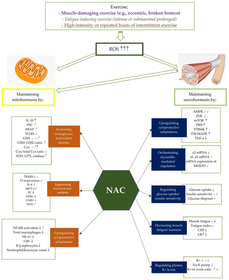
For the hurdles associated with clinical applicability of antioxidants specifically NAC, three major factors might be held responsible i) existence of absence of exhaustive insight with regards to physiological working of oxidative reductive stress. Such kinds of stress reactions are inherent to cellular in addition to organisms feeling of being good , since it possesses the capability of selectively manipulating the working of biomolecules, signal transmission along with physiological working via oxidative reductive modifications. Getting insight with regards to OS is not akin to oxidative injury. Escalating the antioxidant effects capable of stimulating reductive stress , resulting in unanticipated actions. ii) NAC that portrays a thiol possessing compound, possesses the capability to directly decline disulfide bonds/ interfere with their development [54], affecting the protein conformation, their working , as well as their capability of binding to ligands[55,56]. Thereby, apart from its antioxidant characteristics, it might take part in thiol post-translational modifications in addition to affect protein working.iii) The biggest hurdle is the absence of specificity in antioxidant strategies portrays the biggest foundational along with broader challenges. Detecting the optimal dosage poses a big issue as well as an insufficiency of NAC quantities possesses the capability of making the treatment inefficacious. Escalating clarification has been obtained with regards to appropriate controlling of ROS do not prove to be invariably inimical byproducts in addition to work in the form of plethora of indispensable imperative physiological events. ROS along with reactive nitrogen species(RNS ) work in the form of crucial constituents in insulin, MAPK as well as c-Jun-N-terminal kinase (JNK) signaling pathways, possessing part in controlling gene expression, cell growth, differentiation in addition to survival[57]. Figure 2 summarizes the modes by which NAC works(more in exercise rev in ref no 58].
Figure 2
Courtesy ref no -58Potential effects of NAC on mito- and sarcohormesis under exercise-induced oxidative stress. The potential effects of NAC supplementation may vary according to the exercise severity. Therefore, NAC supplementation has been studied on certain exercise intensities including muscle-damaging exercise such as eccentric and broken bronco, fatigue inducing exercise, and high-intensity or repated bouts of intermittent exercise. Each exercise intensity is differently colored and shaped to represent changes in metabolic processes. ↑, ↓, and × represent the alterations caused by muscle damaging exercise (e.g., eccentric, broken bronco) and represent the alterations caused by fatigue-inducing exercise (e.g., intense or submaximal prolonged). ↑, ↓, and × represent the alterations caused by high-intensity or repeated bouts of intermittent exercise. For example, NAC supplementation has showed an enhancing effect of NF-KB activity after fatigue-inducing exercise, while reducing plasma NF-KB activation after muscle-damaging exercise. Abbreviations: NAC: N-acetylcysteine; PC: Protein carbonyls; GSH: reduced glutathione; GPX: glutathione peroxidase; SOD: superoxide dismutase; NADPH: Nicotinamide adenine dinucleotide phosphate; CR: Creatine kinase; GSSG: oxidized glutathione; 3MH: 3-methylhistidine; IL-6: Interleukine-6; TGSH: total glutathione; MCP: Monocyte chemotactic protein; PGC-1a: Peroxisome proliferator-activated receptor coactivator-1a, Cys: cysteine, TBARS: Thiobarbituric acid-reactive substances; PSC: peroxyl radical scavenging capacity; mTOR: polyclonal anti-phospho- mammalian target of rapamycin; p38 MAPK: mitogen activated protein kinase; NF-kB: Nuclear factor kappa B; TNF-a: tumor necrosis factor-a; CRP: C-reactive protein; TAC: total antioxidant capacity; SOD: Superoxide dismutase; FRAP: ferric reducing ability of plasma; CAT: catalase; HLA: human leukocyte antigen; CD11b:(integrin αM); AMPK:AMP activated protein kinase; MDA: malondialdehyde; p70S6K: 70 kDa ribosomal protein S6 kinase, PKB: protein kinase B.
In reference to taking care of the aforementioned three hurdles, the subsequent approaches need to concentrate on exploiting physiological working controlled by oxidative reductive stress. Additionally, they need to guarantee avoidance of extensive antioxidant effects which might lead to oxidative reductive inimicality,all of which takes place,fully taking into account the accurateness needed for redox regulation. Accurateness redox controlling apparently might be the prospective direction in reference to advancing in addition to accurate redox medicine work is currently getting undertaken[59]. Additionally, tackling PCOS by utilization of NAC might work in the form of supplementary approach. The ideal strategy is to escalate physiological working along with attempt regeneration of state by physical exercise , dietary controlling as well as non pharmacological management[60,61].
5. Discussion
Therefore lone NAC as well as NAC in combination with agents, might yield certain advantageous in PCOS treatment .Nonetheless, it is significant to acknowledge that the therapeutic actions of NAC in the circumstances of PCOS are of complicated nature, which continue to be uncharted .An individualized supplementation strategy which implicates customized dosage dependent on separate persons requirement is imperative. Precision biomarkers are required to be isolated in reference to monitoring OS quantities, evaluation of need of NAC supplementation, estimate exact dosage along with asssessment of efficacy of pharmacological management. The maximum efficacious strategy is to endorse. a holistic healthy lifestyle inclusive of harmoneous diet, moderate exercise , as well as commensurate habits, in reference to managing PCOS. Additionally, greater asssessment is needed for attaining insight in reference to the manner NAC works in PCOS women by scientific workers particularly with regards to basic research.
The shortcomings of this review are i) utilization of NAC in combination with other agents( for instance insulin sensitizers, antioxidants in addition to Vitamins) adds complications in evaluating advantageous effects of just lone NAC therapy . A plethora of studies performed have implicated management with utilization of myriad of substances, guaranteeing it to be tough to attribute with clarity that lone NAC is implicated in NAC therapy .ii) physiological part of OS poses a hurdles in attenuating pathological actions of oxidants utilizing antioxidant substances s. Such paradoxical actions is possibly the exposition in view of which certain outcomes obtained utilizing antioxidant substances are disheartening.iii) As per the Rotterdam’s diagnostic criteria PCOS manifestation takes place with variable phenotypes in addition to extra meaning might get added subsequent to contrasting actions of NAC in variable PCOS phenotypes( A/B/C/D subkinds) for illustrating advantageous actions of NAC in variable PCOS patients.
There is need to concentrate on the following points by researchers in further work i) evaluating psychological results subsequent to NAC ingestion in PCOS women ii) estimating the therapeutic dosage required for obtaining the clinically advantageous actions on PCOS characteristics iii) performing systematic reviews as well as and meta-analysis of the publications conducted till date specifically concentrating on the actions of NAC on PCOS characteristics in which case there is paucity of corroboration iv)Use along with documenting standardized diagnostic criteria for PCOS in further studies performed commensurate with the International Guidelines for evaluation as well as management of PCOSv) illustrating further standardized criteria (dosage as well as time period) with inclusion criteria(range of age, BMI in addition to particular PCOS phenotypes) for promoting the contrasting dependable outcomes vi)embracing a greater strategy for monitoring along with sustenance of records with regards to inimical sequelae vii) guaranteeing sufficient powered performed forecoming studies along with paradigmatic population for external corroboration as well as translation in the clinical scenario.
6. Conclusions
As reviewed by us the part of NAC in numerous experimental studies in the form of precautionary treatment or rescue treatment at the time of early stages of sepsis in a rodent model of sepsis stimulated by cecal ligation and puncture(CLP).This is in view of patients with sepsis get usually diagnosed late once there is existence of proven multiorgan impairment,as estimated by sequential organ failure assessment(SOFA)scores[rev in 62]. In this context intravenous treatment with NAC (150mg/kg bolus+20mg/kg/h) in pigs 12hrs subsequent to development of endotoxinemia could not respond with no enhancement in systemic, pulmonary , as well as hepatospanchnic haemodynamics or be correlated with reduction in a biomarker of Oxidative stress(8-isoprostane), inspite of escalation of glutathione amounts[62]. Nevertheless, treatment started at markedly early stage of sepsis are of restricted value with regards to a clinical scenario.Akin outcomes have been displayed in PCOS women with just lesser dosage working whereas failure of greater dosage working. Additionally, we reviewed numerous articles in reference to PCOS pathophysiology, classification in addition to therapy[63-69]. Recently considerable emphasis has been laid in the part of innovative kinds of cell demise for instance ferroptosis along with cuproptosis in case of metabolic disorders for instance non alcoholic fatty liver disease (NAFLD), which might be tackled by utilizing antioxidant Melatonin[70], as well as its part in BC[71].Here we emphasized on part of OS ,oxidative in addition to reductive stress which might be tackled by antioxidant NAC as with role of glutathione(GSH)in ferroptosis in addition to pyroptosis (GSDMD) in PCOS women. Further corroboration was provided by Santander etal.[72 ],in the part of ferroptosis in PCOS where they displayed the existence of lipid peroxidation in women with PCOS, whose modifications occurred subsequent to ingestion of Combined Oral contraceptives (COC), yielding innovative understanding into the pathophysiology of PCOS in the Chilean population[72].
Selenium represents a constituent component of antioxidant enzymes for instance glutathione peroxidase(GPx), which aid in foraging ROS along with diminishing OS. Acknowledged that selenium’s antioxidant characteristics might be advantageous in diminishing oxidative injury, Selenium might possess part in improvement of insulin sensitivity, however not direct hormonal actions [rev in 18]. Furthermore selenium has been recognized to modulate immune reactions, inflammation, as well as influence hormonal balance. Chronic low-grade inflammation is correlated with PCOS, in addition to selenium’s anti-inflammatory actions might aid in better management of PCOS symptoms [rev in 18]. Selenium is a critical constituent of enzymes implicated in the generation in addition to transformation of thyroid hormones [rev in 18]. Thyroid peroxidase (TPO) enzyme is necessary for the generation of thyroid hormones. TPO catalyzes the iodination of tyrosine residues in thyroglobulin, a protein generated by the thyroid gland, which is a crucial step in the production of thyroid hormones, the triiodothyronine (T3) along with thyroxine (T4). Without sufficient selenium, TPO action might be jeopardized, resulting in thyroid hormone dysequilibrium [rev in 18]. In turn, deiodinase enzymes are implicated in transforming one kind of thyroid hormone (T4) into the greater active kind (T3) as well as controlling their accessiblity in variable tissues [rev in 18]. Selenium is required for the appropriate working of these deiodinase enzymes [revin 18]. Certain persons with PCOS might further experience thyroid impairment [rev in 18]. Hormonal dysequilibrium in addition to IR correlated with PCOS are capable of plausiblly influencing thyroid working. Thyroid impairment, specifically situations for instance hypothyroidism, possesses the capability of helping in PCOS symptoms, including irregular menstrual cycles along with hurdles in weight management [rev in 18].
Please view attached pdf to view all references