Thyroid-Like Follicular Carcinoma of the Kidney: A Case Report of a Novel and Emerging Entity
Thyroid-Like Follicular Carcinoma of the Kidney: A Case Report of a Novel and Emerging Entity
Dr Piyali Pramanik *¹, Dr Sanjiban Patra 2, Dr Tarun Jindal3
1. Dr Piyali Pramanik MBBS, MD (Pathology), DipRCPath, Associate Consultant Histopathology, Apollo Multispeciality Hospitals, Kolkata.
2. Dr Sanjiban Patra MBBS (Cal), MD (Pathology), DM (OncoPathology), Senior Consultant OncoPathology, Apollo Multispeciality Hospitals, Kolkata.
3. Dr Tarun Jindal MBBS, MS (Surgery), MCH (Urosurgery), Fellow VUI, DetroitSenior Consultant Uro oncology and Robotic surgery, Apollo Multispeciality Hospitals, Kolkata.
*Correspondence to: Dr Piyali Pramanik MBBS, MD (Pathology), DipRCPath, Associate Consultant Histopathology, Apollo Multispeciality Hospitals, Kolkata.
Copyright.
© 2025 Dr Piyali Pramanik This is an open access article distributed under the Creative Commons Attribution License, which permits unrestricted use, distribution, and reproduction in any medium, provided the original work is properly cited.
Received: 07 Aug 2025
Published: 20 Aug 2025
DOI: https://doi.org/10.5281/zenodo.17283966
Abstract
Thyroid-like follicular carcinoma (TLFC) of the kidney is an extremely rare type of primary renal cell carcinoma, although not been included in the recent WHO classification, but this tumor is considered as a novel and emerging entity in the Genitourinary Pathology Society (GUPS) update. Morphologically the tumor closely resembles follicles of thyroid carcinoma. While the clinical behavior is uncertain due to its rarity, most of them are of low-grade and indolent. In this case report, we document a rare case of TLFC with a short review of the literature to avoid misdiagnosis and add some to the preexisting knowledge.
Keywords: Thyroid-like follicular carcinoma, Immunohistochemistry, Renal cell carcinoma.
Thyroid-Like Follicular Carcinoma of the Kidney: A Case Report of a Novel and Emerging Entity
Abbreviations used:
TLFC Thyroid-like follicular carcinoma;
GUPS Genitourinary Pathology Society;
RCC Renal cell carcinoma;
EMA Epithelial membrane antigen;
CK Cytokeratin;
TTF1 Thyroid transcription factor 1;
Tg Thyroglobulin;
PAX8 Paired homeobox protein 8;
WT1 Wilms tumor antigen 1;
Introduction
Thyroid-like follicular carcinoma of the kidney (TLFC) is an unusual histological type of renal cell carcinoma, which is histologically similar to the primary thyroid follicular or papillary carcinoma. The first case was reported in 2004 and since then, to our knowledge, 40 cases have been reported in the literature.1,2 Here we describe the demographic, radiologic, histopathological and immunohistochemical details of TLFC.
Case History
A 49 year old female patient presented with on and off lower abdominal pain and history of urinary infection. There was no history of hematuria or no relevant family history. Physical examination of the thyroid, abdomen and pelvis was within normal limits. Routine hematological investigations including thyroid function tests, were within normal ranges.
Ultrasound of the abdomen showed a left renal complex cyst measuring 3.5×3.2 cm, which was further clarified on a computed tomography (CT) and Magnetic resonance imaging (MRI)scan describing it as a Bosniak 3 cortical cyst measuring 3.6x3.5x3.1 cm at the upper pole with a small exophytic component, with enhancing thick internal septae. [Fig. 1a] There was no evidence of renal vein involvement, lymphadenopathy, or any metastatic disease. Considering the size and location of the tumor, a robotic partial nephrectomy was performed.
Gross examination of the partial nephrectomy specimen showed a multiloculated cystic lesion measuring 3.6x3.5x3.3 cm with rubbery fibrotic wall and containing thin brownish fluid.
Microscopically, the tumor was relatively well circumscribed, consisting of macro- and micro-follicles of varying sizes filled with amorphous eosinophilic colloid-like material. [Fig 1b] Follicles are lined by cuboidal epithelium with nuclear enlargement, overcrowding, mild clearing and moderate amount of eosinophilic cytoplasm [Fig 1c]. Mitosis and necrosis were absent. Conventional clear cell RCC or other known types of RCCs like areas were not identified. The tumor cells were positive for CK7, PAX8, and Cyclin D1 (Patchy) [Fig 1d-1f] and negative for WT1. Importantly, staining for thyroid transcription factor (TTF-1) and thyroglobulin (Tg) were negative confirming that the tumor was not a metastatic carcinoma of thyroid origin. Hence, based on the morphology and immunohistochemical profile, a diagnosis of TLFC of the kidney [Pathological stage pT1a] without any high grade features. On follow-up, the patient is doing well without any recurrence.
Fig. 1a. MRI showing a renal cortical cyst with enhancing septa (yellow arrow) 1b-1d. Histopathology of the tumor showing thyroid-like follicles containing eosinophilic secretion (H&E, 100-400x) 1e -1f. PAX8 and Cyclin D1 expression in tumor cells respectively (IHC, 400x)
Discussion
Primary TLFCK is a rare and recently described entity.2 This tumor is more common in female (Female: Male = 2:1) with a mean age of 41-44.5 years.1 The main differential diagnoses of the tumor are metastatic thyroid carcinoma, atrophic kidney like lesion and thyroidization in end stage renal disease (ESRD)/ Chronic pyelonephritis. The last one is excluded based on normal clinical profile of the patient and normal urea, creatinine level. Metastasis from the thyroid papillary or follicular carcinoma is excluded by help of immunohistochemistry and normal thyroid profile and thyroid imaging of the patient. TLFCK expresses EMA, CK7 and vimentin. PAX8 has also been reported positive in tumor cells.The most important factor in the reported cases of TLFCK is consistent immuno-negativity for the thyroid-specific markers, such as TTF-1 and Tg. 1
Atrophic kidney, another close differential diagnosis, is positive for WT1 and negative for CK7 and PAX8.
Although the majority of cases reported are low grade with an indolent course, one case in a study showed metastatic extension to the renal hilar lymph nodes and another presented with widespread metastases to the lungs and retroperitoneal lymph nodes.3 One rare incidence of skull and meningeal metastasis was also reported 5 years after the primary diagnosis.5 These cases provide evidence that this rare variant of RCC has a low but distinct malignant potential and can be clinically aggressive.
Surgical resection is the mainstay of treatment. Tumors with a complete capsule, peripheral location and without any metastasis partial nephrectomy or tumor enucleation is sufficient. Tumors > 4?cm or associated with invasive growth or distant metastasis radical nephrectomy, combined with the corresponding regional lymph node dissection is the preferred treatment option.5
This recently described histological variant of renal cell carcinoma, an emerging entity, is important to recognise in order to prevent unnecessary or inappropriate treatment. More data regarding this tumor needs to be collected to ascertain its true clinical behaviour.
Acknowledgement: We acknowledge the entire team in histopathology for their support
Financial support: None
Consent: Proper consent was obtained from the patient for publication of the radiology and histopathology images
Conflict of Interest: The authors have no conflicts of interest.
References
1. Trpkov K, Hes O, Williamson SR, Adeniran AJ, Agaimy A, Alaghehbandan R, et al. New developments in existing WHO entities and evolving molecular concepts: The Genitourinary Pathology Society (GUPS) update on renal neoplasia. Mod Pathol 2021;34(7):1392-1424.
2. Tretiakova MS, Kehr EL, Gore JL, Tykodi SS. Thyroid-Like Follicular Renal Cell Carcinoma Arising Within Benign Mixed Epithelial and Stromal Tumor. Int J Surg Pathol 2020;28(1):80-6.
3. Dhillon J, Tannir NM, Matin SF, Tamboli P, Czerniak BA, Guo CC. Thyroid-like follicular carcinoma of the kidney with metastases to the lungs and retroperitoneal lymph nodes. Hum Pathol 2011;42(1):146-50.
4. Dong L, Huang J, Huang L, Shi O, Liu Q, Chen H, Xue W, Huang Y. Thyroid-Like Follicular Carcinoma of the Kidney in a Patient with Skull and Meningeal Metastasis: A Unique Case Report and Review of the Literature. Medicine 2016;(15):e3314. doi: 10.1097/MD.0000000000003314.
5. Zhang Y, Yang J, Zhang M, Meng Z, Song W, Yang L et al. Thyroid follicular carcinoma-like renal tumor: A case report and literature review. Medicine 2018;97(21):e10815. doi:10.1097/MD.0000000000010815.
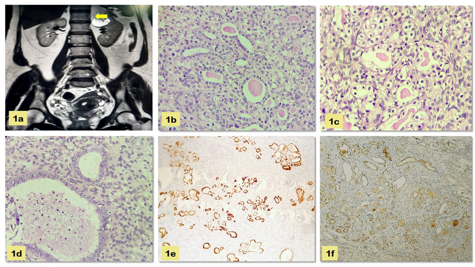
Figure 1